Department of Poisoning and Occupational Diseases, Qilu Hospital, Cheeloo College of Medicine, Shandong University, Jinan, Shandong 250012, P.R. China.
Department of Geriatric Medicine, Qilu Hospital, Cheeloo College of Medicine, Shandong University, Jinan, Shandong 250012, P.R. China.
Mol Med Rep. 2020 Nov;22(5):3687-3694. doi: 10.3892/mmr.2020.11453. Epub 2020 Aug 21.
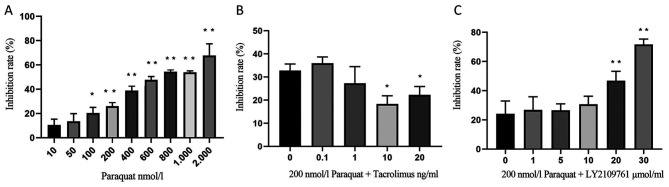
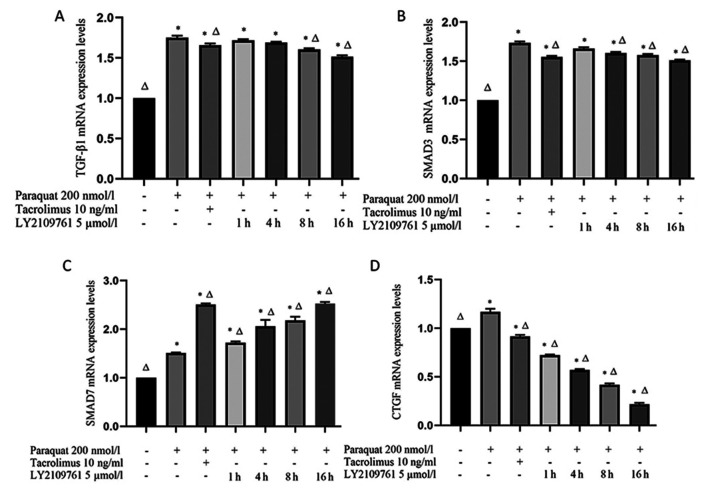
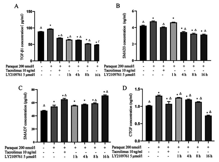
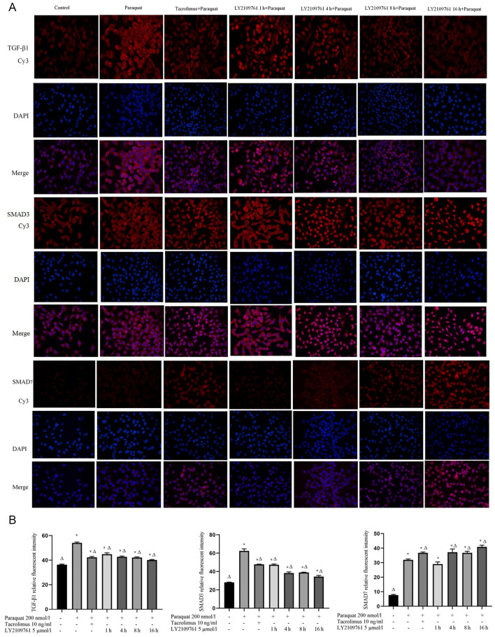
Paraquat is a highly toxic pesticide, which often causes pulmonary interstitial fibrosis after poisoning, and there is no specific antidote. At present, limited studies have reported that tacrolimus, as an immunosuppressant, can inhibit pulmonary fibrosis, but the specific mechanism remains unknown. The aim of the present study was to demonstrate the effect of tacrolimus on the TGF‑β1 pathway associated with pulmonary fibrosis in paraquat exposed alveolar type II epithelial cells, and to identify the antipulmonary fibrosis mechanism of tacrolimus The rat alveolar epithelial type II RLE‑6TN cell line was exposed to paraquat and treated with or without tacrolimus for 24 h, or with a TGF‑β1 receptor type I/II inhibitor (LY2109761) for 1, 4, 8 or 16 h. MTT assays were used to detect the viability of rat alveolar type II epithelial cells under these different treatment conditions, while the concentrations of TGF‑β1, SMAD3, SMAD7 and connective tissue growth factor (CTGF) in the cell culture supernatant were determined using ELISAs. Additionally, reverse transcription‑quantitative PCR and immunofluorescence were used to analyze the mRNA and protein expression levels of TGF‑β1, SMAD3, CTGF and SMAD7. The results demonstrated that the inhibition of the proliferation of RLE‑6TN cells exposed to 200 nmol/l paraquat was 26.05±2.99%. The inhibition rate of 10 ng/ml tacrolimus on paraquat‑exposed alveolar type II epithelial cells was 18.40±3.49%. The inhibition rate caused by 5 µmol/l LY2109761 was 26.56±4.49%. The expression levels of TGF‑β1, SMAD3 and CTGF, as well as their concentrations in the culture supernatant, were significantly downregulated in the tacrolimus group compared with the paraquat group. However, both the concentration and expression levels of SMAD7 were significantly upregulated in the tacrolimus group compared with the paraquat group. In conclusion, tacrolimus can reduce the levels of TGF‑β1, SMAD3 and CTGF, increase the level of SMAD7 in TGF‑β1 signaling pathway and protect the development of pulmonary fibrosis in paraquat exposed alveolar epithelial cells.
百草枯是一种剧毒农药,中毒后常引起肺间质纤维化,目前尚无特效解毒剂。目前有限的研究报道,他克莫司作为一种免疫抑制剂,能抑制肺纤维化,但具体机制尚不清楚。本研究旨在探讨他克莫司对百草枯染毒肺泡Ⅱ型上皮细胞中 TGF-β1 通路的影响,以明确他克莫司抗肺纤维化的作用机制。将大鼠肺泡Ⅱ型上皮细胞系 RLE-6TN 用百草枯染毒,并分别用或不用他克莫司处理 24 h,或用 TGF-β1 受体Ⅰ/Ⅱ型抑制剂(LY2109761)处理 1、4、8 或 16 h。采用 MTT 法检测不同处理条件下大鼠肺泡Ⅱ型上皮细胞的活力,采用 ELISA 法检测细胞培养上清液中 TGF-β1、SMAD3、SMAD7 和结缔组织生长因子(CTGF)的浓度。此外,采用逆转录-定量 PCR 和免疫荧光法分析 TGF-β1、SMAD3、CTGF 和 SMAD7 的 mRNA 和蛋白表达水平。结果表明,浓度为 200 nmol/L 的百草枯抑制 RLE-6TN 细胞增殖率为 26.05%±2.99%。浓度为 10 ng/ml 的他克莫司对百草枯染毒的肺泡Ⅱ型上皮细胞的抑制率为 18.40%±3.49%。浓度为 5 μmol/L 的 LY2109761 的抑制率为 26.56%±4.49%。与百草枯组相比,他克莫司组 TGF-β1、SMAD3 和 CTGF 的表达水平及其培养上清液中的浓度均显著下调,而 SMAD7 的浓度和表达水平均显著上调。结论:他克莫司可降低 TGF-β1、SMAD3 和 CTGF 的水平,增加 TGF-β1 信号通路中 SMAD7 的水平,从而减轻百草枯染毒肺泡上皮细胞肺纤维化的发展。