Department of Ophthalmology, Semmelweis University, Budapest, Hungary.
Department of Neurology, Semmelweis University, Budapest, Hungary.
Geroscience. 2020 Dec;42(6):1499-1525. doi: 10.1007/s11357-020-00252-7. Epub 2020 Oct 4.
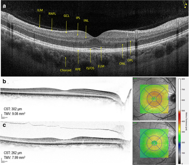
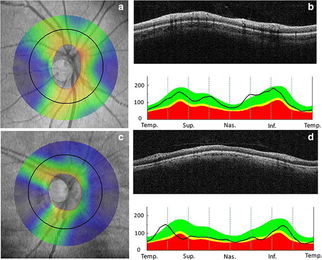
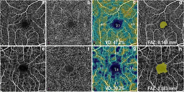
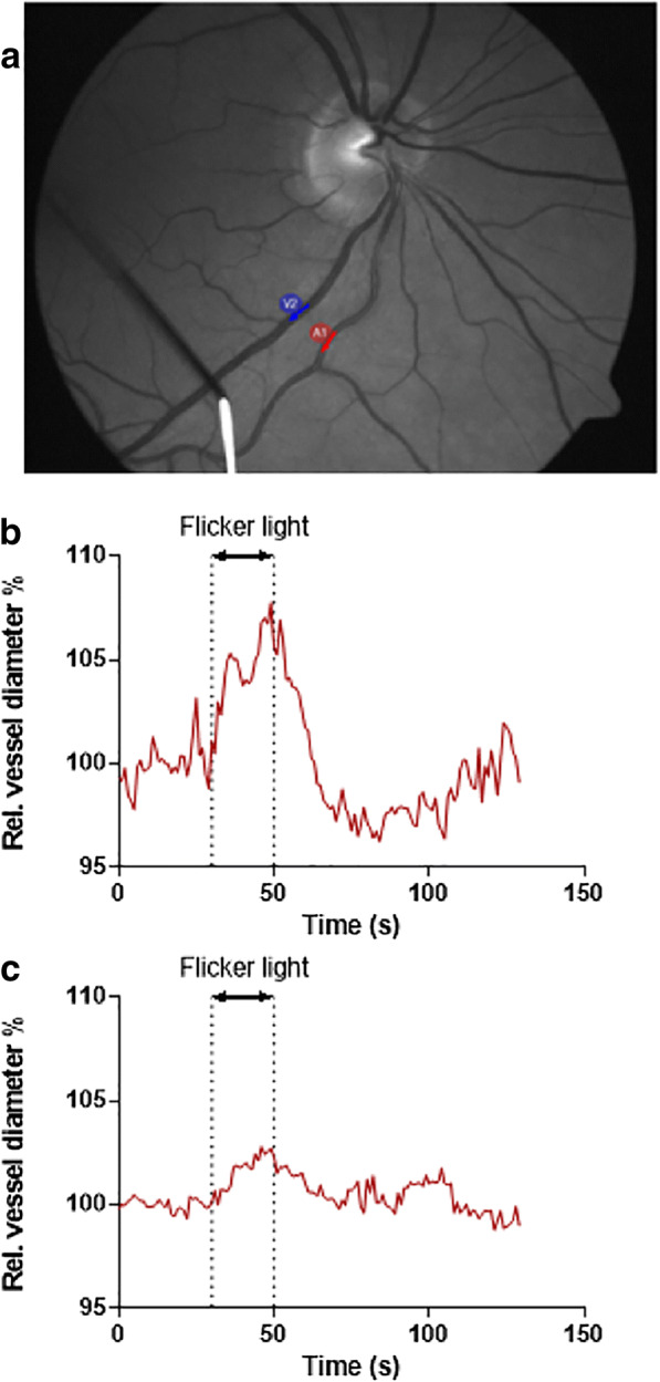
Cognitive impairment and dementia are major medical, social, and economic public health issues worldwide with significant implications for life quality in older adults. The leading causes are Alzheimer's disease (AD) and vascular cognitive impairment/dementia (VCID). In both conditions, pathological alterations of the cerebral microcirculation play a critical pathogenic role. Currently, the main pathological biomarkers of AD-β-amyloid peptide and hyperphosphorylated tau proteins-are detected either through cerebrospinal fluid (CSF) or PET examination. Nevertheless, given that they are invasive and expensive procedures, their availability is limited. Being part of the central nervous system, the retina offers a unique and easy method to study both neurodegenerative disorders and cerebral small vessel diseases in vivo. Over the past few decades, a number of novel approaches in retinal imaging have been developed that may allow physicians and researchers to gain insights into the genesis and progression of cerebromicrovascular pathologies. Optical coherence tomography (OCT), OCT angiography, fundus photography, and dynamic vessel analyzer (DVA) are new imaging methods providing quantitative assessment of retinal structural and vascular indicators-such as thickness of the inner retinal layers, retinal vessel density, foveal avascular zone area, tortuosity and fractal dimension of retinal vessels, and microvascular dysfunction-for cognitive impairment and dementia. Should further studies need to be conducted, these retinal alterations may prove to be useful biomarkers for screening and monitoring dementia progression in clinical routine. In this review, we seek to highlight recent findings and current knowledge regarding the application of retinal biomarkers in dementia assessment.
认知障碍和痴呆是全球范围内主要的医学、社会和经济公共卫生问题,对老年人的生活质量有重大影响。主要病因是阿尔茨海默病(AD)和血管性认知障碍/痴呆(VCID)。在这两种情况下,大脑微循环的病理改变都起着关键的致病作用。目前,AD 的主要病理生物标志物-β-淀粉样肽和过度磷酸化的 tau 蛋白-通过脑脊液(CSF)或 PET 检查来检测。然而,由于这些检查是侵入性和昂贵的,因此其应用受到限制。视网膜作为中枢神经系统的一部分,为研究神经退行性疾病和脑小血管疾病提供了一种独特而简单的方法。在过去的几十年中,已经开发出了许多新的视网膜成像方法,这些方法可能使医生和研究人员能够深入了解脑微血管病变的发生和进展。光学相干断层扫描(OCT)、OCT 血管造影、眼底照相和动态血管分析器(DVA)是新的成像方法,可对视网膜结构和血管指标进行定量评估,如内视网膜层的厚度、视网膜血管密度、黄斑无血管区面积、视网膜血管的迂曲和分形维数以及微血管功能障碍,这些都与认知障碍和痴呆有关。如果需要进一步研究,这些视网膜改变可能被证明是筛查和监测痴呆在临床常规进展的有用生物标志物。在这篇综述中,我们旨在强调视网膜生物标志物在痴呆评估中的应用的最新发现和现有知识。