Wang Kankai, Ru Junnan, Zhang Hengli, Chen Jiayu, Lin Xiao, Lin Zhongxiao, Wen Min, Huang Lijie, Ni Haoqi, Zhuge Qichuan, Yang Su
Zhejiang Provincial Key Laboratory of Aging and Neurological Disorder Research, The First Affiliated Hospital of Wenzhou Medical University, Wenzhou, China.
Department of Neurosurgery, The First Affiliated Hospital of Wenzhou Medical University, Wenzhou, China.
Front Neurosci. 2020 Aug 18;14:848. doi: 10.3389/fnins.2020.00848. eCollection 2020.
Ischemic stroke-induced inflammation and inflammasome-dependent pyroptotic neural death cause serious neurological injury. Nano-sized plasma exosomes have exhibited therapeutic potential against ischemia and reperfusion injury by ameliorating inflammation. To enhance its therapeutic potential in patients with ischemic injury, we isolated exosomes from melatonin-treated rat plasma and assessed the neurological protective effect in a rat model of focal cerebral ischemia.
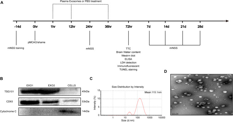
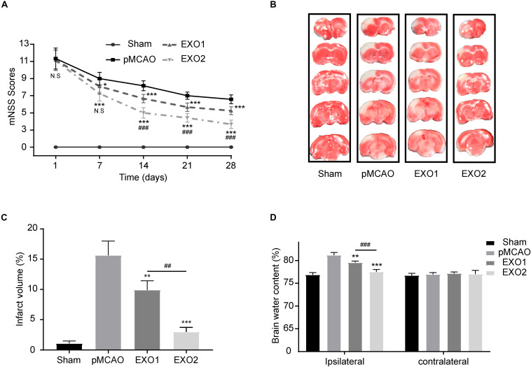
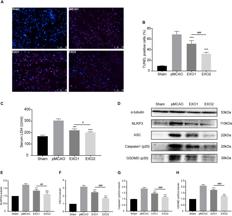
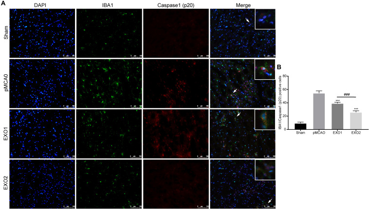
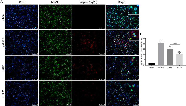
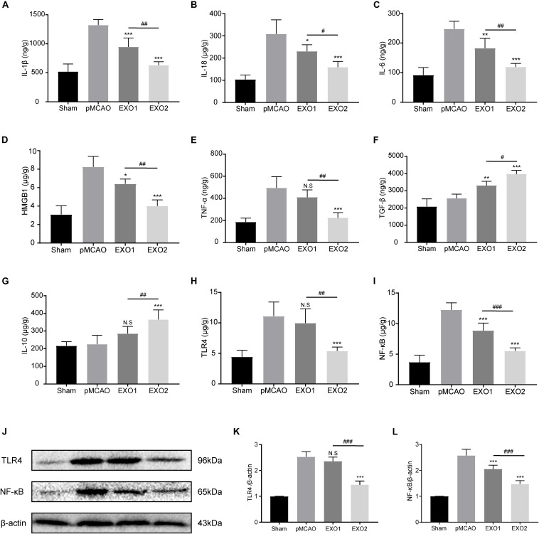
Basal plasma exosomes and melatonin-treated plasma exosomes were isolated and intravenously injected into a rat model of focal cerebral ischemia. Neurological recovery was evaluated by determining the modified neurological severity score (mNSS), infarct volume, and brain water content. Pyroptosis in the ischemic cortex was detected through dUTP nick-end labeling (TUNEL) assay, lactate dehydrogenase (LDH) release, and gasdermin D (GSDMD) cleavage. NLRP3 inflammasome assembly and global inflammatory cytokine secretion were detected by enzyme-linked immunosorbent assay (ELISA) and Western blot assay. In immunized Sprague-Dawley rats, microglia pyroptosis was determined through a positive percentage of IBA1 and caspase-1 (p20) cells. Finally, the microRNA (miRNA) profiles in melatonin-treated plasma exosomes were analyzed by exosome miRNA microarray analysis.
Melatonin treatment enhanced plasma exosome therapeutic effects against ischemia-induced inflammatory responses and inflammasome-mediated pyroptosis. In addition, we confirmed that ischemic stroke-induced pyroptotic cell death occurred in the microglia and neuron, while the administration of melatonin-treated exosomes further effectively decreased the infarct volume and improved recovery of function regulation of the TLR4/NF-κB signaling pathway. Finally, the altered miRNA profiles in the melatonin-treated plasma exosomes demonstrated the regulatory mechanisms involved in neurological recovery after ischemic injury.
This study suggests that nano-sized plasma exosomes with melatonin pretreatment might be a more effective strategy for patients with ischemic brain injury. Further exploration of key molecules in the plasma exosome may provide increased therapeutic value for cerebral ischemic injury.
缺血性中风引发的炎症以及炎性小体依赖性细胞焦亡导致的神经死亡会造成严重的神经损伤。纳米级血浆外泌体已通过减轻炎症展现出对缺血再灌注损伤的治疗潜力。为增强其对缺血性损伤患者的治疗潜力,我们从褪黑素处理的大鼠血浆中分离出外泌体,并在局灶性脑缺血大鼠模型中评估其神经保护作用。
分离基础血浆外泌体和褪黑素处理的血浆外泌体,并将其静脉注射到局灶性脑缺血大鼠模型中。通过测定改良神经功能缺损评分(mNSS)、梗死体积和脑含水量来评估神经功能恢复情况。通过脱氧尿苷三磷酸缺口末端标记(TUNEL)检测、乳酸脱氢酶(LDH)释放和gasdermin D(GSDMD)裂解来检测缺血皮质中的细胞焦亡。通过酶联免疫吸附测定(ELISA)和蛋白质印迹法检测NLRP3炎性小体组装和整体炎性细胞因子分泌。在免疫的Sprague-Dawley大鼠中,通过IBA1和半胱天冬酶-1(p20)细胞的阳性百分比来确定小胶质细胞焦亡。最后,通过外泌体微小RNA(miRNA)微阵列分析来分析褪黑素处理的血浆外泌体中的miRNA谱。
褪黑素处理增强了血浆外泌体对缺血诱导的炎症反应和炎性小体介导的细胞焦亡的治疗效果。此外,我们证实缺血性中风诱导的细胞焦亡性细胞死亡发生在小胶质细胞和神经元中,而给予褪黑素处理的外泌体进一步有效降低了梗死体积并改善了功能恢复,这是通过TLR4/NF-κB信号通路的调节实现的。最后,褪黑素处理的血浆外泌体中改变的miRNA谱证明了缺血性损伤后神经功能恢复所涉及的调节机制。
本研究表明,经褪黑素预处理的纳米级血浆外泌体可能是缺血性脑损伤患者更有效的治疗策略。进一步探索血浆外泌体中的关键分子可能会为脑缺血损伤提供更高的治疗价值。