Yasuda Ippei, Shima Tomoko, Moriya Taiki, Ikebuchi Ryoyo, Kusumoto Yutaka, Ushijima Akemi, Nakashima Akitoshi, Tomura Michio, Saito Shigeru
Department of Obstetrics and Gynecology, University of Toyama, Toyama, Japan.
Laboratory of Immunology, Faculty of Pharmacy, Osaka Ohtani University, Osaka, Japan.
Front Immunol. 2020 Sep 11;11:557720. doi: 10.3389/fimmu.2020.557720. eCollection 2020.
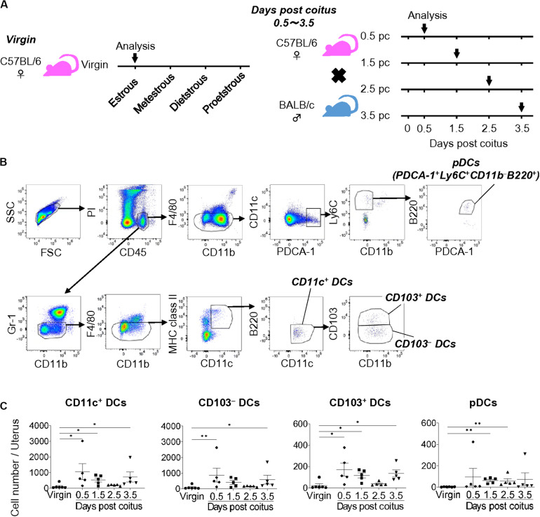
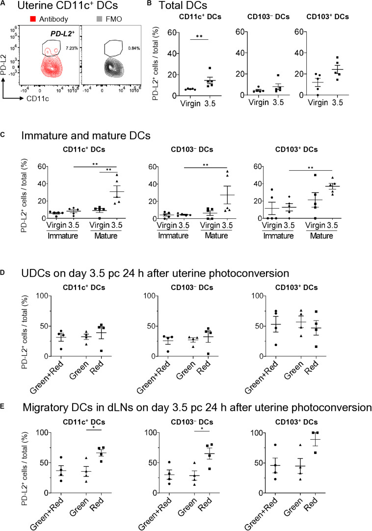
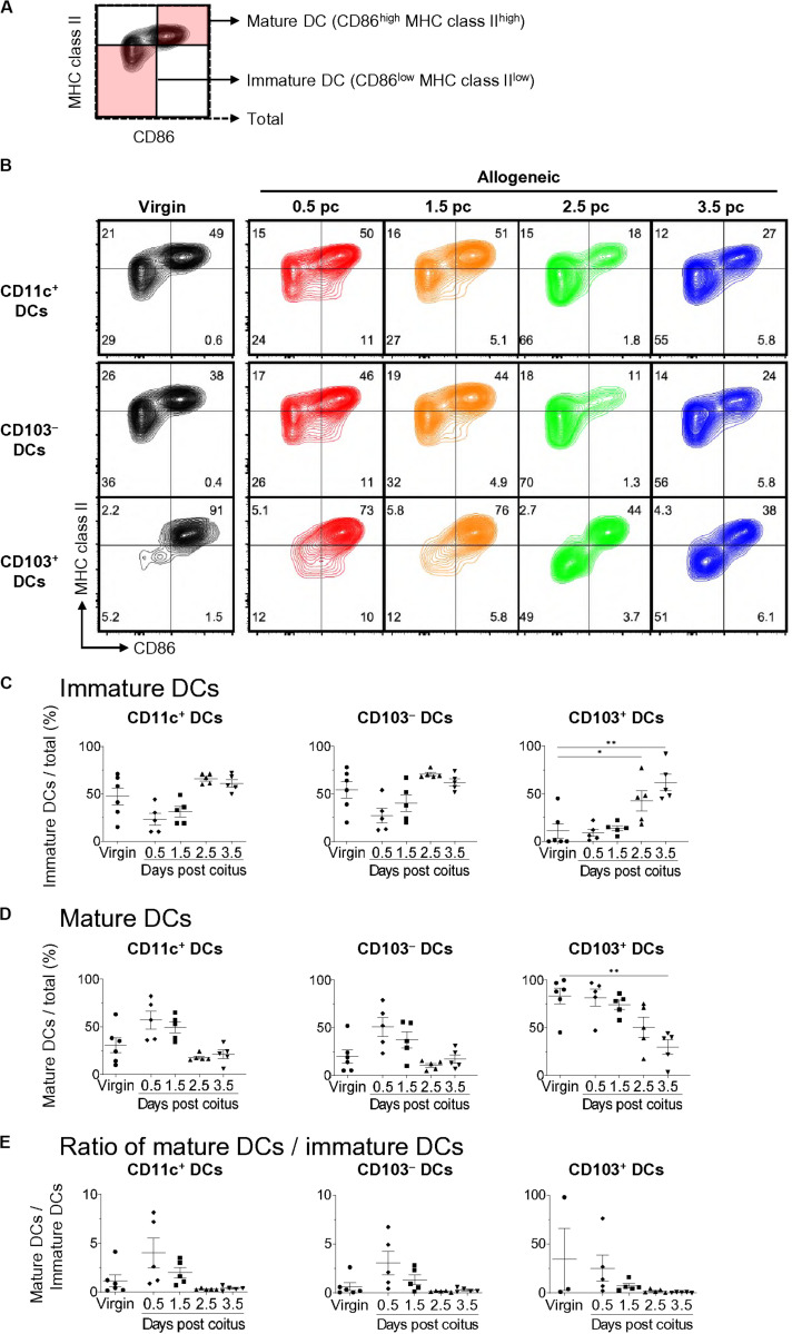
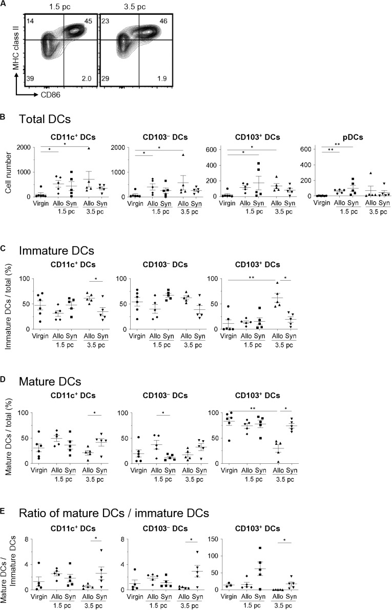
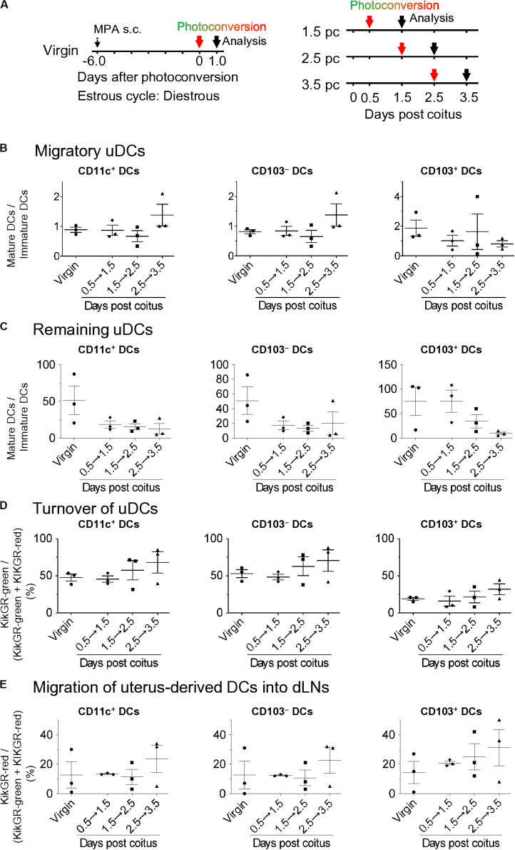
Dendritic cells (DCs) are essential for successful embryo implantation. However, the properties of uterine DCs (uDCs) during the implantation period are not well characterized. In this study, we investigated the dynamic changes in the uDC phenotypes during the period between coitus and implantation. In virgin mice, we evaluated the expressions of CD103 and XCR1, this is the first report to demonstrate uDCs expressing CD103 in XCR1cDC1s and XCR1cDC2s. On day 0.5 post coitus (pc), the number of uterine CD11cCD103MHC classIICD86-mature DCs rapidly increased and then decreased to non-pregnancy levels on days 1.5 and 2.5 pc. On day 3.5 pc just before implantation, the number of CD11cCD103MHC class IICD86-immature DCs increased in the uterus. The increase in mature uDCs on day 1.5 pc was observed in both allogeneic- and syngeneic mating, suggesting that sexual intercourse, or semen, play a role in this process. Meanwhile, the increase in immature uDCs on day 3.5 pc was only observed in allogeneic mating, suggesting that allo-antigens in the semen contribute to this process. Next, to understand the turnover and migration of uDCs, we monitored DC movement in the uterus and uterine draining lymph nodes (dLNs) using photoconvertible protein Kikume Green Red (KikGR) mice. On day 0.5 pc, uDCs were composed of equal numbers of remaining DCs and migratory DCs. However, on day 3.5 pc, uDCs were primarily composed of migratory DCs, suggesting that most of the uDCs migrate from the periphery just before implantation. Finally, we studied the expression of PD-L2-which induces immunoregulation-on DCs. On day 3.5 pc, PD-L2 was expressed on CD103-mature and CD103-mature DCs in the uterus. However, PD-L2 expression on CD103-immature DCs and CD103-immature DCs was very low. Furthermore, both remaining and migratory DCs in the uterus and uterus-derived-DCs in the dLNs on day 3.5 pc highly expressed PD-L2 on their surface. Therefore, our study findings provide a better understanding of the dynamic changes occurring in uterine DCs and dLNs in preparation for implantation following allogeneic- and syngeneic mating.
树突状细胞(DCs)对于胚胎成功着床至关重要。然而,着床期子宫树突状细胞(uDCs)的特性尚未得到充分表征。在本研究中,我们调查了交配至着床期间uDC表型的动态变化。在未交配的小鼠中,我们评估了CD103和XCR1的表达,这是首次报道在XCR1cDC1s和XCR1cDC2s中表达CD103的uDCs。在交配后第0.5天(pc),子宫中CD11cCD103MHC II类CD86 -成熟DCs的数量迅速增加,然后在交配后第1.5天和2.5天降至非妊娠水平。在着床前的第3.5天pc,子宫中CD11cCD103MHC IICD86 -未成熟DCs的数量增加。在第1.5天pc时成熟uDCs的增加在同种异体交配和同基因交配中均观察到,这表明性交或精液在这一过程中起作用。同时,在第3.5天pc时未成熟uDCs的增加仅在同种异体交配中观察到,这表明精液中的同种异体抗原促成了这一过程。接下来,为了了解uDCs的更新和迁移,我们使用光转换蛋白Kikume Green Red(KikGR)小鼠监测子宫和子宫引流淋巴结(dLNs)中的DC运动。在第0.5天pc时,uDCs由数量相等的残留DCs和迁移DCs组成。然而,在第3.5天pc时,uDCs主要由迁移DCs组成,这表明大多数uDCs在着床前从外周迁移而来。最后,我们研究了诱导免疫调节的PD - L2在DCs上的表达。在第3.5天pc时,PD - L2在子宫中CD103 -成熟和CD103 -成熟DCs上表达。然而,PD - L2在CD103 -未成熟DCs和CD103 -未成熟DCs上的表达非常低。此外,在第3.5天pc时,子宫中的残留和迁移DCs以及dLNs中源自子宫的DCs在其表面均高表达PD - L2。因此,我们的研究结果有助于更好地理解同种异体交配和同基因交配后子宫DCs和dLNs为着床所发生的动态变化。