• 文献检索
  • 文档翻译
  • 深度研究
  • 学术资讯
  • Suppr Zotero 插件Zotero 插件
  • 邀请有礼
  • 套餐&价格
  • 历史记录
应用&插件
Suppr Zotero 插件Zotero 插件浏览器插件Mac 客户端Windows 客户端微信小程序
定价
高级版会员购买积分包购买API积分包
服务
文献检索文档翻译深度研究API 文档MCP 服务
关于我们
关于 Suppr公司介绍联系我们用户协议隐私条款
关注我们

Suppr 超能文献

核心技术专利:CN118964589B侵权必究
粤ICP备2023148730 号-1Suppr @ 2026

文献检索

告别复杂PubMed语法,用中文像聊天一样搜索,搜遍4000万医学文献。AI智能推荐,让科研检索更轻松。

立即免费搜索

文件翻译

保留排版,准确专业,支持PDF/Word/PPT等文件格式,支持 12+语言互译。

免费翻译文档

深度研究

AI帮你快速写综述,25分钟生成高质量综述,智能提取关键信息,辅助科研写作。

立即免费体验

基于矢状位脂肪抑制和横轴位T2加权磁共振成像的纹理分析用于确定直肠癌的局部侵犯

Texture Analysis Based on Sagittal Fat-Suppression and Transverse T2-Weighted Magnetic Resonance Imaging for Determining Local Invasion of Rectal Cancer.

作者信息

Lu H C, Wang F, Yin J D

机构信息

School of Medicine and Bioinformatics Engineering, Northeastern University, Shenyang, China.

Department of Radiology, Shengjing Hospital of China Medical University, Shenyang, China.

出版信息

Front Oncol. 2020 Aug 18;10:1476. doi: 10.3389/fonc.2020.01476. eCollection 2020.

DOI:10.3389/fonc.2020.01476
PMID:33014786
原文链接:https://pmc.ncbi.nlm.nih.gov/articles/PMC7461892/
Abstract

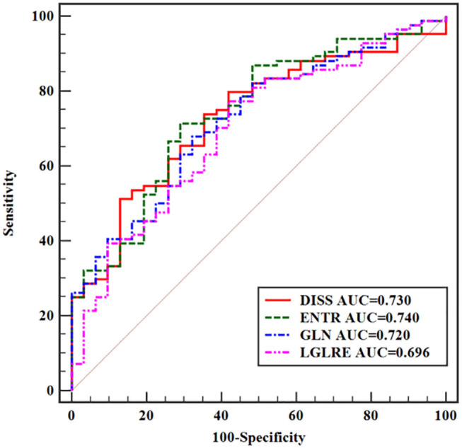
Accurate evaluation of local invasion (T-stage) of rectal cancer is essential for treatment planning. A search of PubMed database indicated that the correlation between texture features from T2-weighted magnetic resonance imaging (T2WI) (MRI) and T-stage has not been explored extensively. To evaluate the performance of texture analysis using sagittal fat-suppression combined with transverse T2WI for determining T-stage of rectal cancer. One hundred and seventy-four rectal cancer cases who underwent preoperative MRI were retrospectively selected and divided into high (T3/4) and low (T1/2) T-stage groups. Texture features were, respectively, extracted from sagittal fat-suppression and transverse T2WI images. Univariate and multivariate analyses were conducted to determine T-stage. Discrimination performance was assessed by receiver operating characteristic (ROC) analysis. For univariate analysis, the best performance in differentiating T1/2 from T3/4 tumors was achieved from transverse T2WI, and the area under the ROC curve (AUC) was 0.740. For multivariate analysis, the logical regression model incorporating the independent predictors achieved an AUC of 0.789. Texture features from sagittal fat-suppression combined with transverse T2WI presented moderate association with T-stage of rectal cancer. These findings may be valuable in selecting optimum treatment strategy.

摘要

准确评估直肠癌的局部侵犯情况(T分期)对于治疗方案的制定至关重要。对PubMed数据库的检索表明,T2加权磁共振成像(T2WI)(MRI)的纹理特征与T分期之间的相关性尚未得到广泛研究。为了评估矢状位脂肪抑制联合横轴位T2WI纹理分析在确定直肠癌T分期中的性能。回顾性选取174例术前行MRI检查的直肠癌病例,分为高(T3/4)、低(T1/2)T分期组。分别从矢状位脂肪抑制和横轴位T2WI图像中提取纹理特征。进行单因素和多因素分析以确定T分期。通过受试者操作特征(ROC)分析评估鉴别性能。单因素分析中,在区分T1/2与T3/4肿瘤方面,横轴位T2WI表现最佳,ROC曲线下面积(AUC)为0.740。多因素分析中,纳入独立预测因素的逻辑回归模型的AUC为0.789。矢状位脂肪抑制联合横轴位T2WI的纹理特征与直肠癌T分期呈中度相关。这些发现可能对选择最佳治疗策略具有重要价值。

https://cdn.ncbi.nlm.nih.gov/pmc/blobs/71d9/7461892/e629b2140f3b/fonc-10-01476-g0005.jpg
https://cdn.ncbi.nlm.nih.gov/pmc/blobs/71d9/7461892/9165469ee54b/fonc-10-01476-g0001.jpg
https://cdn.ncbi.nlm.nih.gov/pmc/blobs/71d9/7461892/e99dc525a1d7/fonc-10-01476-g0002.jpg
https://cdn.ncbi.nlm.nih.gov/pmc/blobs/71d9/7461892/11cefc2fb2b7/fonc-10-01476-g0003.jpg
https://cdn.ncbi.nlm.nih.gov/pmc/blobs/71d9/7461892/ba2a6d657828/fonc-10-01476-g0004.jpg
https://cdn.ncbi.nlm.nih.gov/pmc/blobs/71d9/7461892/e629b2140f3b/fonc-10-01476-g0005.jpg

相似文献

1
Texture Analysis Based on Sagittal Fat-Suppression and Transverse T2-Weighted Magnetic Resonance Imaging for Determining Local Invasion of Rectal Cancer.基于矢状位脂肪抑制和横轴位T2加权磁共振成像的纹理分析用于确定直肠癌的局部侵犯
Front Oncol. 2020 Aug 18;10:1476. doi: 10.3389/fonc.2020.01476. eCollection 2020.
2
Application of Texture Analysis Based on Sagittal Fat-Suppression and Oblique Axial T2-Weighted Magnetic Resonance Imaging to Identify Lymph Node Invasion Status of Rectal Cancer.基于矢状位脂肪抑制和斜轴位T2加权磁共振成像的纹理分析在直肠癌淋巴结侵犯状态识别中的应用
Front Oncol. 2020 Aug 7;10:1364. doi: 10.3389/fonc.2020.01364. eCollection 2020.
3
Performances of Whole Tumor Texture Analysis Based on MRI: Predicting Preoperative T Stage of Rectal Carcinomas.基于磁共振成像的全肿瘤纹理分析性能:预测直肠癌术前T分期
Front Oncol. 2021 Aug 3;11:678441. doi: 10.3389/fonc.2021.678441. eCollection 2021.
4
[Application value of texture analysis of magnetic resonance images in prediction of neoadjuvant chemoradiotherapy efficacy for rectal cancer].磁共振图像纹理分析在预测直肠癌新辅助放化疗疗效中的应用价值
Zhonghua Wei Chang Wai Ke Za Zhi. 2018 Sep 25;21(9):1051-1058.
5
Whole-Tumor Histogram and Texture Imaging Features on Magnetic Resonance Imaging Combined With Epstein-Barr Virus Status to Predict Disease Progression in Patients With Nasopharyngeal Carcinoma.磁共振成像上的全肿瘤直方图和纹理成像特征联合爱泼斯坦-巴尔病毒状态预测鼻咽癌患者的疾病进展
Front Oncol. 2021 Mar 9;11:610804. doi: 10.3389/fonc.2021.610804. eCollection 2021.
6
MRI-based texture analysis of the primary tumor for pre-treatment prediction of bone metastases in prostate cancer.基于MRI的原发性肿瘤纹理分析用于前列腺癌骨转移的治疗前预测
Magn Reson Imaging. 2019 Jul;60:76-84. doi: 10.1016/j.mri.2019.03.007. Epub 2019 Mar 24.
7
Clinical value of T2-weighted imaging combined with diffusion-weighted imaging in preoperative T staging of urinary bladder cancer: a large-scale, multiobserver prospective study on 3.0-T MRI.T2 加权成像联合扩散加权成像对膀胱癌术前 T 分期的临床价值:3.0-T MRI 多中心前瞻性大样本研究。
Acad Radiol. 2013 Aug;20(8):939-46. doi: 10.1016/j.acra.2013.02.012. Epub 2013 Jun 5.
8
Combining quantitative and qualitative magnetic resonance imaging features to differentiate anorectal malignant melanoma from low rectal cancer.结合定量和定性磁共振成像特征以鉴别肛管直肠恶性黑色素瘤与低位直肠癌。
Precis Clin Med. 2021 Apr 30;4(2):119-128. doi: 10.1093/pcmedi/pbab011. eCollection 2021 Jun.
9
T staging of rectal cancer: accuracy of diffusion-weighted imaging compared with T2-weighted imaging on 3.0 tesla MRI.直肠癌 T 分期:3.0T 磁共振弥散加权成像与 T2 加权成像的准确性比较。
J Dig Dis. 2014 Apr;15(4):188-94. doi: 10.1111/1751-2980.12124.
10
MRI-based radiomics of rectal cancer: preoperative assessment of the pathological features.基于 MRI 的直肠癌放射组学:术前病理特征评估。
BMC Med Imaging. 2019 Nov 12;19(1):86. doi: 10.1186/s12880-019-0392-7.

引用本文的文献

1
Artificial intelligence and radiomics in magnetic resonance imaging of rectal cancer: a review.人工智能与放射组学在直肠癌磁共振成像中的应用综述
Explor Target Antitumor Ther. 2023;4(3):406-421. doi: 10.37349/etat.2023.00142. Epub 2023 Jun 30.
2
Virtual biopsy in abdominal pathology: where do we stand?腹部病理学中的虚拟活检:我们目前的进展如何?
BJR Open. 2023 Feb 28;5(1):20220055. doi: 10.1259/bjro.20220055. eCollection 2023.
3
Infliximab response associates with radiologic findings in bio-naïve Crohn's disease.英夫利昔单抗应答与生物初治克罗恩病的影像学发现相关。

本文引用的文献

1
Prediction of Clinical Pathologic Prognostic Factors for Rectal Adenocarcinoma: Volumetric Texture Analysis Based on Apparent Diffusion Coefficient Maps.基于表观扩散系数图的直肠腺癌临床病理预后因素的预测:容积纹理分析。
J Med Syst. 2019 Nov 7;43(12):331. doi: 10.1007/s10916-019-1464-5.
2
Rectal cancer: can T2WI histogram of the primary tumor help predict the existence of lymph node metastasis?直肠癌:原发肿瘤 T2WI 直方图能否帮助预测淋巴结转移的存在?
Eur Radiol. 2019 Dec;29(12):6469-6476. doi: 10.1007/s00330-019-06328-z. Epub 2019 Jul 5.
3
Magnetic Resonance Texture Analysis in Identifying Complete Pathological Response to Neoadjuvant Treatment in Locally Advanced Rectal Cancer.
Eur Radiol. 2023 Aug;33(8):5247-5257. doi: 10.1007/s00330-023-09542-y. Epub 2023 Mar 16.
4
Combining Clinicopathology, IVIM-DWI and Texture Parameters for a Nomogram to Predict Treatment Response to Neoadjuvant Chemoradiotherapy in Locally Advanced Rectal Cancer Patients.结合临床病理学、体素内不相干运动扩散加权成像(IVIM-DWI)和纹理参数构建列线图以预测局部晚期直肠癌患者新辅助放化疗的治疗反应
Front Oncol. 2022 May 27;12:886101. doi: 10.3389/fonc.2022.886101. eCollection 2022.
5
Texture Analysis in the Assessment of Rectal Cancer: Comparison of T2WI and Diffusion-Weighted Imaging.纹理分析在直肠癌评估中的应用:T2WI 与弥散加权成像的比较。
Comput Math Methods Med. 2021 Sep 15;2021:9976440. doi: 10.1155/2021/9976440. eCollection 2021.
6
Radiomics and machine learning applications in rectal cancer: Current update and future perspectives.直肠癌的放射组学和机器学习应用:现状更新与未来展望。
World J Gastroenterol. 2021 Aug 28;27(32):5306-5321. doi: 10.3748/wjg.v27.i32.5306.
7
Radiomics and Magnetic Resonance Imaging of Rectal Cancer: From Engineering to Clinical Practice.直肠癌的放射组学与磁共振成像:从工程到临床实践
Diagnostics (Basel). 2021 Apr 23;11(5):756. doi: 10.3390/diagnostics11050756.
磁共振纹理分析在识别局部晚期直肠癌新辅助治疗的完全病理缓解中的应用
Dis Colon Rectum. 2019 Feb;62(2):163-170. doi: 10.1097/DCR.0000000000001224.
4
Preoperative radiomic signature based on multiparametric magnetic resonance imaging for noninvasive evaluation of biological characteristics in rectal cancer.基于多参数磁共振成像的术前放射组学特征用于非侵入性评估直肠癌的生物学特征。
Eur Radiol. 2019 Jun;29(6):3200-3209. doi: 10.1007/s00330-018-5763-x. Epub 2018 Nov 9.
5
MRI radiomics analysis for predicting preoperative synchronous distant metastasis in patients with rectal cancer.MRI 放射组学分析预测直肠癌患者术前同步远处转移
Eur Radiol. 2019 Aug;29(8):4418-4426. doi: 10.1007/s00330-018-5802-7. Epub 2018 Nov 9.
6
Role of texture analysis in breast MRI as a cancer biomarker: A review.纹理分析在乳腺 MRI 作为癌症生物标志物中的作用:综述。
J Magn Reson Imaging. 2019 Apr;49(4):927-938. doi: 10.1002/jmri.26556. Epub 2018 Nov 3.
7
Role of Quantitative Dynamic Contrast-Enhanced MRI in Evaluating Regional Lymph Nodes With a Short-Axis Diameter of Less Than 5 mm in Rectal Cancer.定量动态对比增强 MRI 在评估直肠癌短轴直径小于 5mm 的区域淋巴结中的作用。
AJR Am J Roentgenol. 2019 Jan;212(1):77-83. doi: 10.2214/AJR.18.19866. Epub 2018 Oct 24.
8
Preoperative tumor texture analysis on MRI predicts high-risk disease and reduced survival in endometrial cancer.术前磁共振肿瘤纹理分析预测子宫内膜癌的高危疾病和降低生存率。
J Magn Reson Imaging. 2018 Dec;48(6):1637-1647. doi: 10.1002/jmri.26184. Epub 2018 Aug 13.
9
Rectal cancer: ESMO Clinical Practice Guidelines for diagnosis, treatment and follow-up.直肠癌:欧洲肿瘤内科学会临床实践指南之诊断、治疗及随访
Ann Oncol. 2017 Jul 1;28(suppl_4):iv22-iv40. doi: 10.1093/annonc/mdx224.
10
Endometrial Carcinoma: MR Imaging-based Texture Model for Preoperative Risk Stratification-A Preliminary Analysis.子宫内膜癌:基于磁共振成像的纹理模型用于术前风险分层——初步分析。
Radiology. 2017 Sep;284(3):748-757. doi: 10.1148/radiol.2017161950. Epub 2017 May 10.