Department of Multidisciplinary Regenerative Research, Guglielmo Marconi University, Via Vittoria Colonna, 11, 00193 Rome, Italy.
San Rossore Dental Unit, Viale delle Cascine 152 San Rossore, 56122 Pisa, Italy.
Int J Environ Res Public Health. 2020 Oct 1;17(19):7199. doi: 10.3390/ijerph17197199.
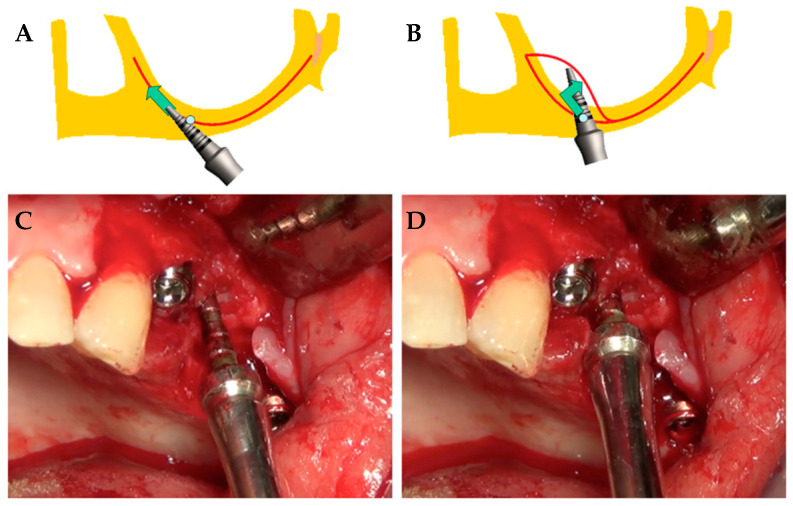
The present study is designed to compare the outcomes of two sinus augmentation procedures: distal displacement of the anterior wall versus standard sinus lifting and grafting with a lateral window approach.
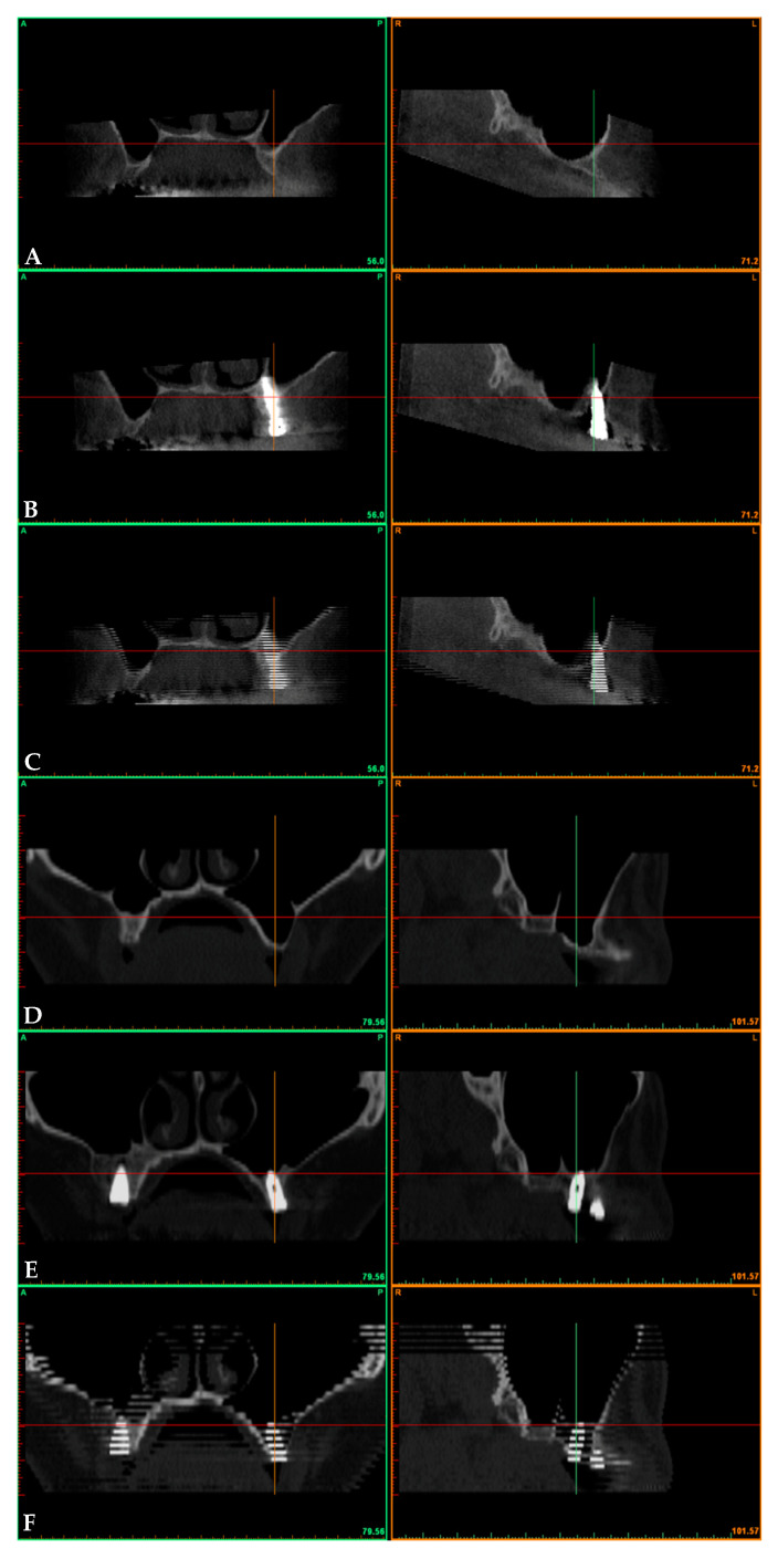
In the displacement group, a localized surgical fracture of the sinus floor achieved through an electromagnetic device results in the distal displacement of the anterior wall. In the filling group, sinus lifting (with lateral access) and grafting with particulate xenogeneic bone substitute was performed. Bone volume beneath the maxillary sinus was investigated with computerized tomography after baseline and postoperative data superimposition. Clinical and radiological outcomes over three years had been evaluated.
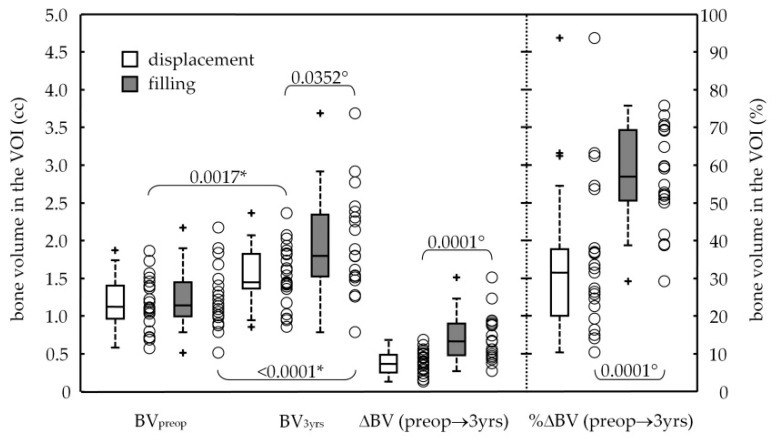
Forty-three dental implants were selected. The two sinus lift procedures significantly increased the bone volume (-value ≤ 0.0017) in the displacement group from 1.17 ± 0.34 to 1.53 ± 0.39 cc, with a final bone gain of +0.36 ± 0.17 cc, and in the filling group from 1.24 ± 0.41 to 1.94 ± 0.68 cc, with a bone augmentation of +0.71 ± 0.31 cc. No events of dental implant bulging into the maxillary sinus occurred. Two implants failed early on in the filling group, attesting the 3-year survival rate of 92.6% (CI95%: 82.7-100%). Marginal bone loss at the distal aspect was 1.66 ± 0.72 and 1.25 ± 0.78 mm, respectively, for the displacement and filling groups, with a significant difference (-value = 0.0497).
Results showed a significant and effective bone gain around dental implants at a 3-year survey for both sinus augmented by backward displacement of the anterior wall (+34%) and sinus lifting and grafting with a lateral window approach (+57%).
本研究旨在比较两种窦提升术的结果:前壁远移与标准窦提升和外侧壁开窗植骨。
在移位组中,通过电磁设备实现的鼻窦地板局部手术骨折导致前壁远移。在填充组中,进行了鼻窦提升(外侧入路)和颗粒异种骨替代物的植骨。在基线和术后数据叠加后,使用计算机断层扫描(CT)检查上颌窦下方的骨量。在三年内评估了临床和放射学结果。
选择了 43 颗牙种植体。两种窦提升术均显著增加了移位组的骨量(-值≤0.0017),从 1.17±0.34 增加到 1.53±0.39cc,最终骨增量为+0.36±0.17cc,在填充组中,从 1.24±0.41 增加到 1.94±0.68cc,骨增量为+0.71±0.31cc。没有牙种植体膨入上颌窦的事件发生。在填充组中,有两个种植体早期失败,证明了 3 年的存活率为 92.6%(95%CI:82.7-100%)。远侧边缘骨丢失分别为 1.66±0.72 和 1.25±0.78mm,移位和填充组之间存在显著差异(-值=0.0497)。
结果显示,在 3 年的调查中,通过后移前壁增加窦(增加 34%)和通过外侧壁开窗提升和植骨增加窦(增加 57%),两种方法都能显著有效地增加牙种植体周围的骨量。