• 文献检索
  • 文档翻译
  • 深度研究
  • 学术资讯
  • Suppr Zotero 插件Zotero 插件
  • 邀请有礼
  • 套餐&价格
  • 历史记录
应用&插件
Suppr Zotero 插件Zotero 插件浏览器插件Mac 客户端Windows 客户端微信小程序
定价
高级版会员购买积分包购买API积分包
服务
文献检索文档翻译深度研究API 文档MCP 服务
关于我们
关于 Suppr公司介绍联系我们用户协议隐私条款
关注我们

Suppr 超能文献

核心技术专利:CN118964589B侵权必究
粤ICP备2023148730 号-1Suppr @ 2026

文献检索

告别复杂PubMed语法,用中文像聊天一样搜索,搜遍4000万医学文献。AI智能推荐,让科研检索更轻松。

立即免费搜索

文件翻译

保留排版,准确专业,支持PDF/Word/PPT等文件格式,支持 12+语言互译。

免费翻译文档

深度研究

AI帮你快速写综述,25分钟生成高质量综述,智能提取关键信息,辅助科研写作。

立即免费体验

未经染色的人体皮肤活检的 3d 相衬体层析成像术可能可以识别出不同个体的真皮和表皮之间的形态差异。

3d phase-contrast nanotomography of unstained human skin biopsies may identify morphological differences in the dermis and epidermis between subjects.

机构信息

Institute for X-Ray Physics, University of Göttingen, Göttingen, Germany.

Cluster of Excellence "Multiscale Bioimaging: from Molecular Machines to Networks of Excitable Cells" (MBExC), University of Göttingen, Göttingen, Germany.

出版信息

Skin Res Technol. 2021 May;27(3):316-323. doi: 10.1111/srt.12974. Epub 2020 Oct 6.

DOI:10.1111/srt.12974
PMID:33022848
原文链接:https://pmc.ncbi.nlm.nih.gov/articles/PMC8246570/
Abstract

BACKGROUND

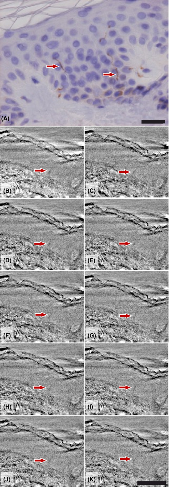
Enteric neuropathy is described in most patients with gastrointestinal dysmotility and may be found together with reduced intraepidermal nerve fiber density (IENFD). The aim of this pilot study was to assess whether three-dimensional (3d) imaging of skin biopsies could be used to examine various tissue components in patients with gastrointestinal dysmotility.

MATERIAL AND METHODS

Four dysmotility patients of different etiology and two healthy volunteers were included. From each subject, two 3-mm punch skin biopsies were stained with antibodies against protein gene product 9.5 or evaluated as a whole with two X-ray phase-contrast computed tomography (CT) setups, a laboratory µCT setup and a dedicated synchrotron radiation nanoCT end-station.

RESULTS

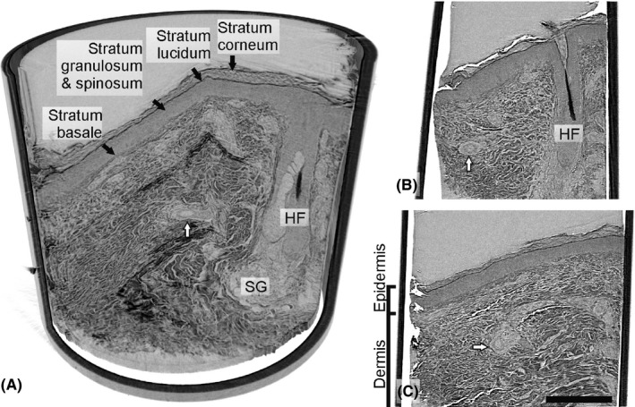
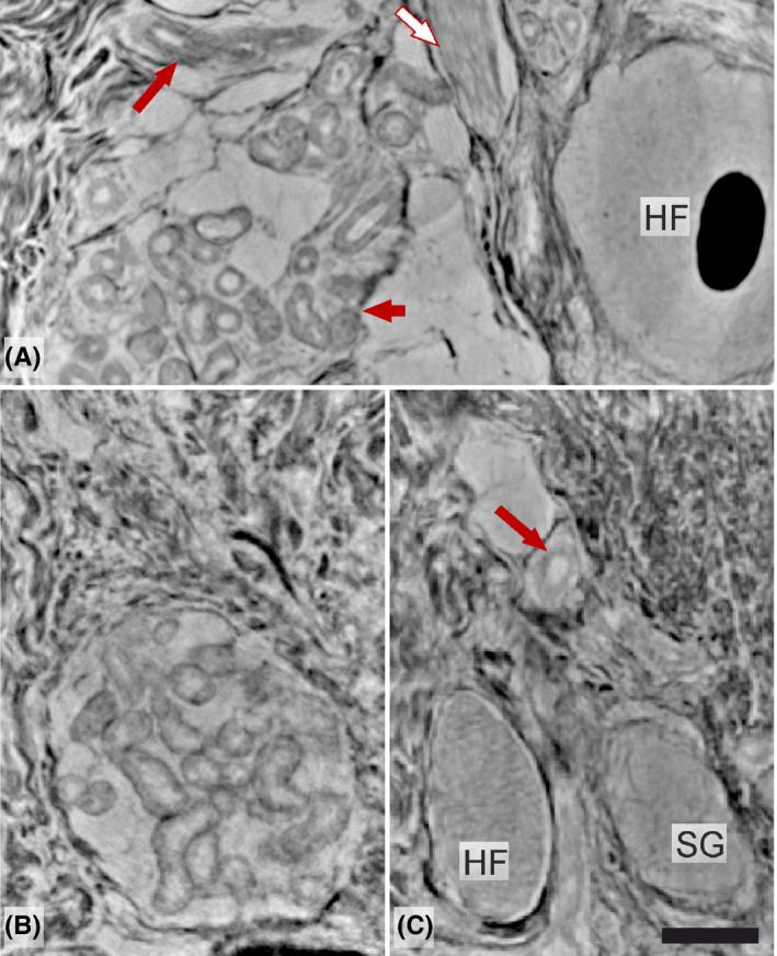
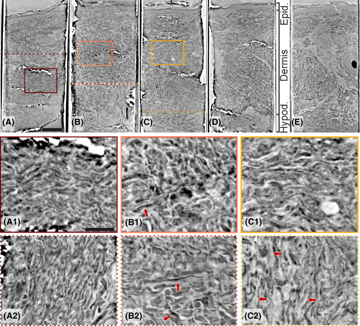
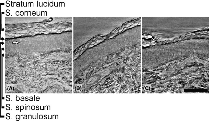
Two patients had reduced IENFD, and two normal IENFD, compared with controls. µCT and X-ray phase-contrast holographic nanotomography scanned whole tissue specimens, with optional high-resolution scans revealing delicate structures, without differentiation of various fibers and cells. Irregular architecture of dermal fibers was observed in the patient with Ehlers-Danlos syndrome and the patient with idiopathic dysmotility showed an abundance of mesenchymal ground substance.

CONCLUSIONS

3d phase-contrast tomographic imaging may be useful to illustrate traits of connective tissue dysfunction in various organs and to demonstrate whether disorganized dermal fibers could explain organ dysfunction.

摘要

背景

肠神经病在大多数胃肠道动力障碍患者中均有描述,并且可能与表皮内神经纤维密度降低(IENFD)一起发现。本初步研究的目的是评估三维(3d)成像皮肤活检是否可用于检查胃肠道动力障碍患者的各种组织成分。

材料和方法

纳入了四位不同病因的动力障碍患者和两位健康志愿者。从每位受试者中,取两个 3mm 打孔皮肤活检,用针对蛋白基因产物 9.5 的抗体染色,或用两种 X 射线相衬 CT(CT)设置、实验室 µCT 设置和专用同步辐射纳米 CT 端站进行整体评估。

结果

与对照组相比,有两位患者的 IENFD 降低,有两位患者的 IENFD 正常。µCT 和 X 射线相衬全息纳米断层扫描可扫描整个组织标本,可选的高分辨率扫描可揭示细微结构,而无需区分各种纤维和细胞。在埃勒斯-当洛斯综合征患者中观察到真皮纤维的不规则结构,而特发性动力障碍患者表现出丰富的间质基质。

结论

3d 相衬断层成像可能有助于说明各种器官中结缔组织功能障碍的特征,并证明紊乱的真皮纤维是否可以解释器官功能障碍。

https://cdn.ncbi.nlm.nih.gov/pmc/blobs/5907/8246570/2c41daeac28c/SRT-27-316-g002.jpg
https://cdn.ncbi.nlm.nih.gov/pmc/blobs/5907/8246570/22deb996dd0e/SRT-27-316-g001.jpg
https://cdn.ncbi.nlm.nih.gov/pmc/blobs/5907/8246570/e0ed1547292a/SRT-27-316-g003.jpg
https://cdn.ncbi.nlm.nih.gov/pmc/blobs/5907/8246570/2afd3f982288/SRT-27-316-g004.jpg
https://cdn.ncbi.nlm.nih.gov/pmc/blobs/5907/8246570/e71dae7fb71d/SRT-27-316-g005.jpg
https://cdn.ncbi.nlm.nih.gov/pmc/blobs/5907/8246570/2c41daeac28c/SRT-27-316-g002.jpg

相似文献

1
3d phase-contrast nanotomography of unstained human skin biopsies may identify morphological differences in the dermis and epidermis between subjects.未经染色的人体皮肤活检的 3d 相衬体层析成像术可能可以识别出不同个体的真皮和表皮之间的形态差异。
Skin Res Technol. 2021 May;27(3):316-323. doi: 10.1111/srt.12974. Epub 2020 Oct 6.
2
Three-dimensional imaging of nociceptive intraepidermal nerve fibers in human skin biopsies.人皮肤活检中伤害性表皮内神经纤维的三维成像。
J Vis Exp. 2013 Apr 29(74):e50331. doi: 10.3791/50331.
3
Subepidermal Schwann cell counts correlate with skin innervation - an exploratory study.表皮下施万细胞计数与皮肤神经支配相关——一项探索性研究。
Muscle Nerve. 2022 Apr;65(4):471-479. doi: 10.1002/mus.27496. Epub 2022 Jan 26.
4
Quantitative and qualitative normative dataset for intraepidermal nerve fibers using skin biopsy.使用皮肤活检的表皮内神经纤维定量和定性标准数据集。
PLoS One. 2018 Jan 25;13(1):e0191614. doi: 10.1371/journal.pone.0191614. eCollection 2018.
5
The reliability of skin biopsy with measurement of intraepidermal nerve fiber density.通过测量表皮内神经纤维密度进行皮肤活检的可靠性。
J Neurol Sci. 2005 Jan 15;228(1):65-9. doi: 10.1016/j.jns.2004.09.032. Epub 2004 Nov 5.
6
Reduction of Intraepidermal Nerve Fiber Density (IENFD) in the skin biopsies of patients with fibromyalgia: a controlled study.纤维肌痛患者皮肤活检中表皮内神经纤维密度(IENFD)的降低:一项对照研究。
J Neurol Sci. 2014 Dec 15;347(1-2):143-7. doi: 10.1016/j.jns.2014.09.035. Epub 2014 Sep 28.
7
Intraepidermal Nerve Fiber Density in Postmortem Skin: A Novel Approach.
J Clin Neuromuscul Dis. 2019 Dec;21(2):69-76. doi: 10.1097/CND.0000000000000264.
8
Morphological Characterisation of Unstained and Intact Tissue Micro-architecture by X-ray Computed Micro- and Nano-Tomography.通过X射线计算机微纳断层扫描对未染色完整组织微结构进行形态学表征
Sci Rep. 2015 May 15;5:10074. doi: 10.1038/srep10074.
9
Evaluation of small nerve fiber dysfunction in type 2 diabetes.评估 2 型糖尿病中小纤维神经功能障碍。
Acta Neurol Scand. 2020 Jan;141(1):38-46. doi: 10.1111/ane.13171. Epub 2019 Oct 9.
10
Epidermal nerve fiber density: normative reference range and diagnostic efficiency.表皮神经纤维密度:正常参考范围及诊断效能。
Arch Neurol. 1998 Dec;55(12):1513-20. doi: 10.1001/archneur.55.12.1513.

引用本文的文献

1
Structure of the myenteric plexus in normal and diseased human ileum analyzed by X-ray virtual histology slices.X 射线虚拟组织切片分析正常和患病人类回肠的肌间神经丛结构。
World J Gastroenterol. 2022 Aug 7;28(29):3994-4006. doi: 10.3748/wjg.v28.i29.3994.

本文引用的文献

1
A phase-retrieval toolbox for X-ray holography and tomography.用于X射线全息术和断层扫描的相位恢复工具箱。
J Synchrotron Radiat. 2020 May 1;27(Pt 3):852-859. doi: 10.1107/S1600577520002398. Epub 2020 Apr 14.
2
Three-dimensional architecture of human diabetic peripheral nerves revealed by X-ray phase contrast holographic nanotomography.X 射线相衬全息纳米断层成像术揭示的人糖尿病外周神经的三维结构。
Sci Rep. 2020 May 5;10(1):7592. doi: 10.1038/s41598-020-64430-5.
3
Phase-contrast x-ray tomography of neuronal tissue at laboratory sources with submicron resolution.
实验室光源下具有亚微米分辨率的神经元组织相衬X射线断层扫描术。
J Med Imaging (Bellingham). 2020 Jan;7(1):013502. doi: 10.1117/1.JMI.7.1.013502. Epub 2020 Feb 20.
4
Autonomic and peripheral neuropathy with reduced intraepidermal nerve fiber density can be observed in patients with gastrointestinal dysmotility.在胃肠动力障碍患者中可观察到自主神经和周围神经病变伴表皮内神经纤维密度降低。
Clin Case Rep. 2019 Dec 4;8(1):142-148. doi: 10.1002/ccr3.2575. eCollection 2020 Jan.
5
Spatial relationship between telocytes, interstitial cells of Cajal and the enteric nervous system in the human ileum and colon.人回肠和结肠中 telocytes、Cajal 间质细胞与肠神经系统之间的空间关系。
J Cell Mol Med. 2020 Mar;24(6):3399-3406. doi: 10.1111/jcmm.15013. Epub 2020 Jan 26.
6
Absence of Collagen Flowers on Electron Microscopy and Identification of (Likely) Pathogenic Variants in Two Patients.电子显微镜下未见胶原花,两名患者中存在(可能)致病性变异体。
Genes (Basel). 2019 Sep 27;10(10):762. doi: 10.3390/genes10100762.
7
Evaluation of small nerve fiber dysfunction in type 2 diabetes.评估 2 型糖尿病中小纤维神经功能障碍。
Acta Neurol Scand. 2020 Jan;141(1):38-46. doi: 10.1111/ane.13171. Epub 2019 Oct 9.
8
X-ray-Based 3D Virtual Histology-Adding the Next Dimension to Histological Analysis.基于 X 射线的 3D 虚拟组织学——为组织学分析增添新维度。
Mol Imaging Biol. 2018 Oct;20(5):732-741. doi: 10.1007/s11307-018-1246-3.
9
Hard X-Ray Nanoholotomography: Large-Scale, Label-Free, 3D Neuroimaging beyond Optical Limit.硬X射线纳米全息断层成像:超越光学极限的大规模、无标记三维神经成像
Adv Sci (Weinh). 2018 Mar 30;5(6):1700694. doi: 10.1002/advs.201700694. eCollection 2018 Jun.
10
Three-dimensional virtual histology of human cerebellum by X-ray phase-contrast tomography.基于 X 射线相衬断层成像术的人类小脑三维虚拟组织学
Proc Natl Acad Sci U S A. 2018 Jul 3;115(27):6940-6945. doi: 10.1073/pnas.1801678115. Epub 2018 Jun 18.