Institute for X-Ray Physics, University of Göttingen, 37077 Göttingen, Germany.
Center for Nanoscopy and Molecular Physiology of the Brain, 37073 Göttingen, Germany.
Proc Natl Acad Sci U S A. 2018 Jul 3;115(27):6940-6945. doi: 10.1073/pnas.1801678115. Epub 2018 Jun 18.
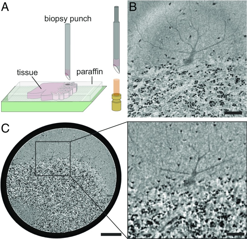
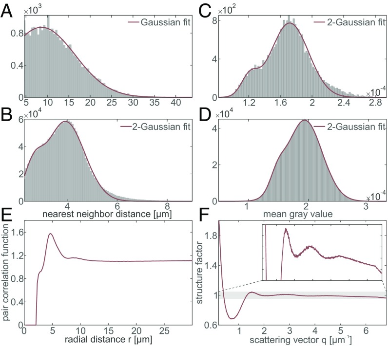
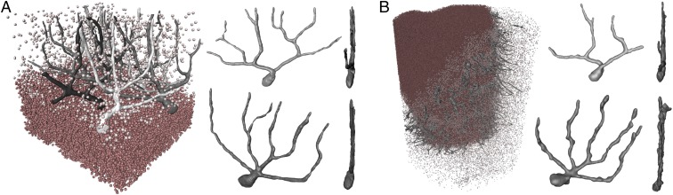
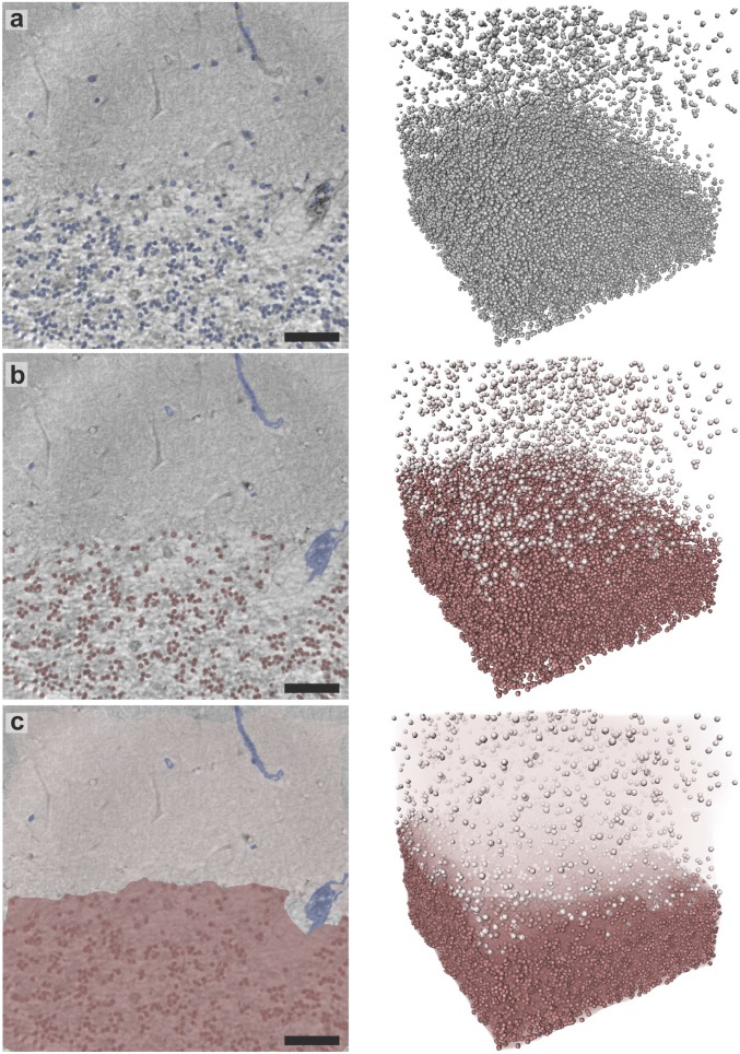
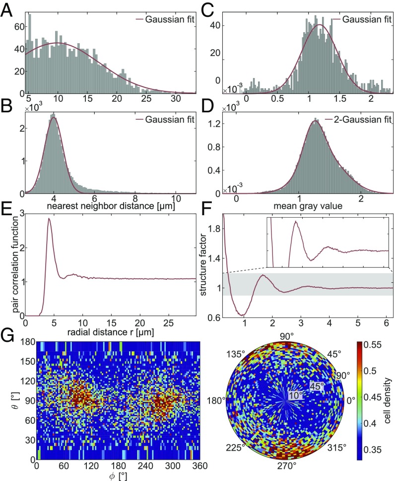
To quantitatively evaluate brain tissue and its corresponding function, knowledge of the 3D cellular distribution is essential. The gold standard to obtain this information is histology, a destructive and labor-intensive technique where the specimen is sliced and examined under a light microscope, providing 3D information at nonisotropic resolution. To overcome the limitations of conventional histology, we use phase-contrast X-ray tomography with optimized optics, reconstruction, and image analysis, both at a dedicated synchrotron radiation endstation, which we have equipped with X-ray waveguide optics for coherence and wavefront filtering, and at a compact laboratory source. As a proof-of-concept demonstration we probe the 3D cytoarchitecture in millimeter-sized punches of unstained human cerebellum embedded in paraffin and show that isotropic subcellular resolution can be reached at both setups throughout the specimen. To enable a quantitative analysis of the reconstructed data, we demonstrate automatic cell segmentation and localization of over 1 million neurons within the cerebellar cortex. This allows for the analysis of the spatial organization and correlation of cells in all dimensions by borrowing concepts from condensed-matter physics, indicating a strong short-range order and local clustering of the cells in the granular layer. By quantification of 3D neuronal "packing," we can hence shed light on how the human cerebellum accommodates 80% of the total neurons in the brain in only 10% of its volume. In addition, we show that the distribution of neighboring neurons in the granular layer is anisotropic with respect to the Purkinje cell dendrites.
为了定量评估脑组织及其相应功能,了解 3D 细胞分布是必不可少的。获取此信息的金标准是组织学,这是一种破坏性和劳动密集型技术,其中标本被切片并在光学显微镜下检查,以非各向同性的分辨率提供 3D 信息。为了克服传统组织学的局限性,我们使用具有优化光学、重建和图像分析的相衬 X 射线断层扫描,分别在专用同步辐射端站和紧凑型实验室源进行,我们在该端站配备了 X 射线波导光学器件,用于相干和波前滤波。作为概念验证演示,我们探测了未经染色的人类小脑毫米大小的蜡块中的 3D 细胞结构,并表明在两个设置中都可以在整个标本中达到各向同性的亚细胞分辨率。为了能够对重建数据进行定量分析,我们演示了在小脑皮层内自动分割和定位超过 100 万个神经元。这允许通过借用凝聚态物理学的概念来分析所有维度的细胞的空间组织和相关性,表明颗粒层中的细胞具有强烈的短程有序性和局部聚类。通过对 3D 神经元“包装”的量化,我们可以了解人类小脑如何在其体积的 10%中容纳大脑中 80%的总神经元。此外,我们还表明,颗粒层中相邻神经元的分布相对于浦肯野细胞树突是各向异性的。