Department of Radiology, Mayo Clinic, 200 First St SW, Rochester, MN 55905, USA.
Department of Health Sciences Research, Mayo Clinic, 200 First St SW, Rochester, MN 55905, USA.
Neuroimage. 2021 Jan 1;224:117433. doi: 10.1016/j.neuroimage.2020.117433. Epub 2020 Oct 6.
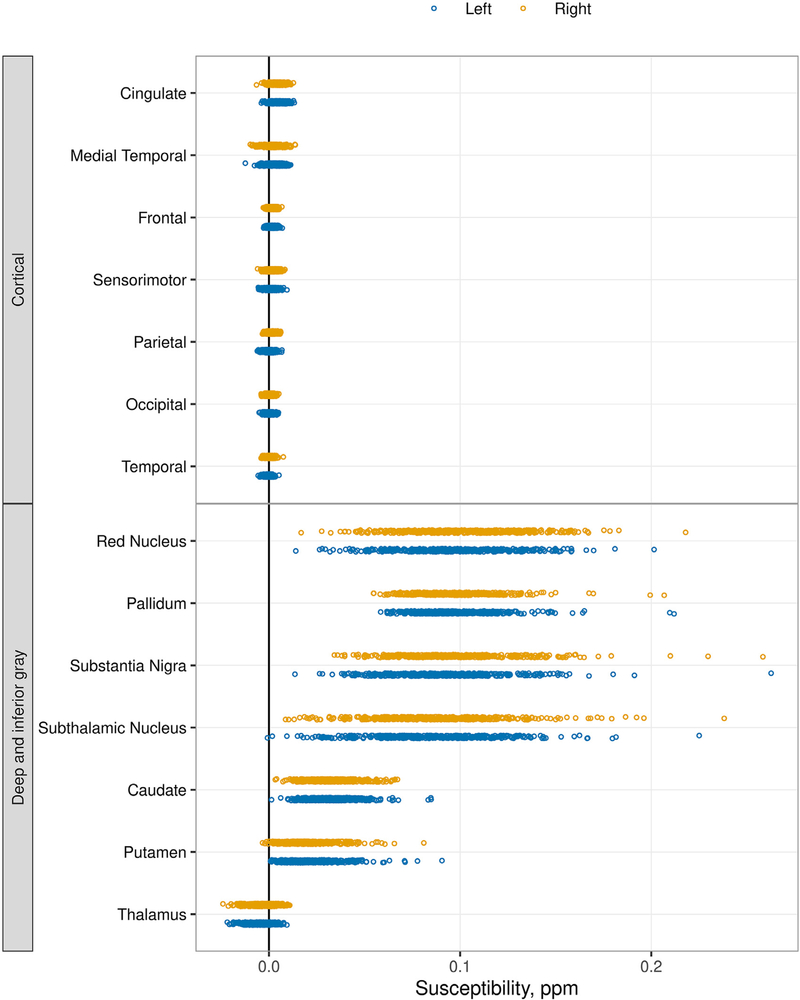
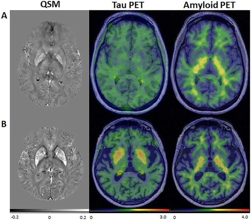
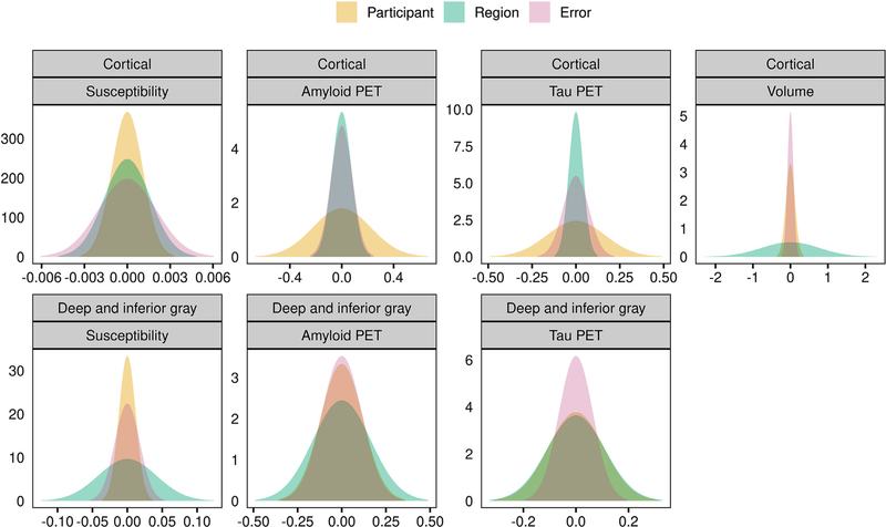
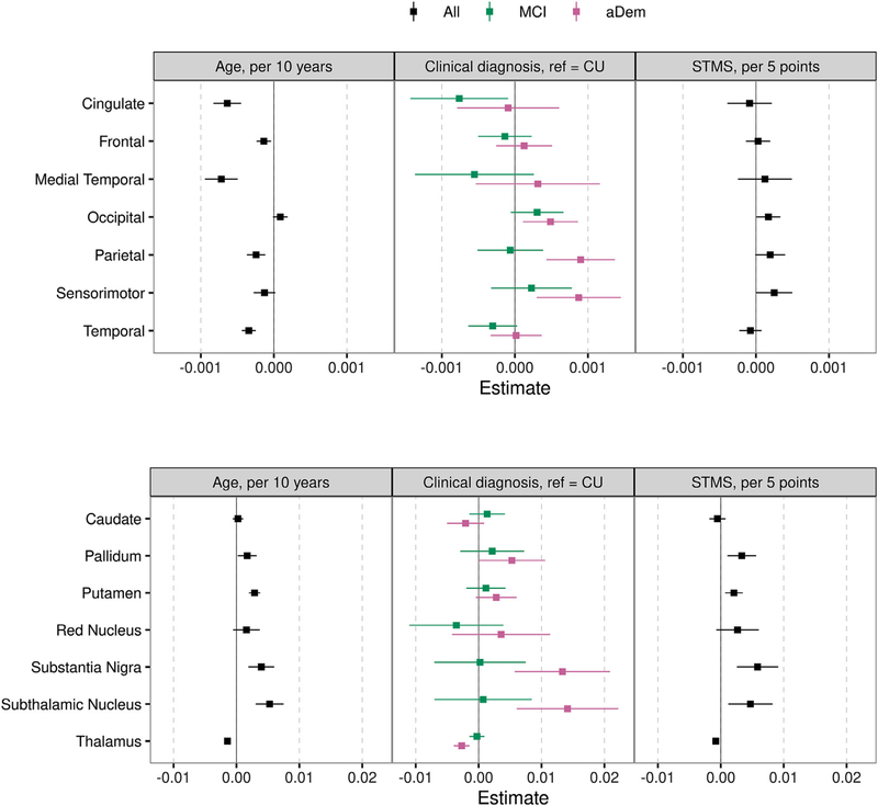
Altered iron metabolism has been hypothesized to be associated with Alzheimer's disease pathology, and prior work has shown associations between iron load and beta amyloid plaques. Quantitative susceptibility mapping (QSM) is a recently popularized MR technique to infer local tissue susceptibility secondary to the presence of iron as well as other minerals. Greater QSM values imply greater iron concentration in tissue. QSM has been used to study relationships between cerebral iron load and established markers of Alzheimer's disease, however relationships remain unclear. In this work we study QSM signal characteristics and associations between susceptibility measured on QSM and established clinical and imaging markers of Alzheimer's disease. The study included 421 participants (234 male, median age 70 years, range 34-97 years) from the Mayo Clinic Study of Aging and Alzheimer's Disease Research Center; 296 (70%) had a diagnosis of cognitively unimpaired, 69 (16%) mild cognitive impairment, and 56 (13%) amnestic dementia. All participants had multi-echo gradient recalled echo imaging, PiB amyloid PET, and Tauvid tau PET. Variance components analysis showed that variation in cortical susceptibility across participants was low. Linear regression models were fit to assess associations with regional susceptibility. Expected increases in susceptibility were found with older age and cognitive impairment in the deep and inferior gray nuclei (pallidum, putamen, substantia nigra, subthalamic nucleus) (betas: 0.0017 to 0.0053 ppm for a 10 year increase in age, p = 0.03 to <0.001; betas: 0.0021 to 0.0058 ppm for a 5 point decrease in Short Test of Mental Status, p = 0.003 to p<0.001). Effect sizes in cortical regions were smaller, and the age associations were generally negative. Higher susceptibility was significantly associated with higher amyloid PET SUVR in the pallidum and putamen (betas: 0.0029 and 0.0012 ppm for a 20% increase in amyloid PET, p = 0.05 and 0.02, respectively), higher tau PET in the basal ganglia with the largest effect size in the pallidum (0.0082 ppm for a 20% increase in tau PET, p<0.001), and with lower cortical gray matter volume in the medial temporal lobe (0.0006 ppm for a 20% decrease in volume, p = 0.03). Overall, these findings suggest that susceptibility in the deep and inferior gray nuclei, particularly the pallidum and putamen, may be a marker of cognitive decline, amyloid deposition, and off-target binding of the tau ligand. Although iron has been demonstrated in amyloid plaques and in association with neurodegeneration, it is of insufficient quantity to be reliably detected in the cortex using this implementation of QSM.
铁代谢的改变与阿尔茨海默病的病理有关,先前的研究表明铁负荷与β淀粉样斑块之间存在关联。定量磁化率映射(QSM)是一种最近流行的磁共振技术,可以推断由于铁以及其他矿物质的存在而导致的局部组织磁化率。更高的 QSM 值意味着组织中铁浓度更高。QSM 已被用于研究大脑铁负荷与阿尔茨海默病的既定标志物之间的关系,但关系仍不清楚。在这项工作中,我们研究了 QSM 信号特征以及 QSM 上测量的磁化率与阿尔茨海默病的既定临床和影像学标志物之间的关系。该研究包括来自梅奥诊所衰老研究和阿尔茨海默病研究中心的 421 名参与者(234 名男性,中位年龄 70 岁,范围 34-97 岁);296 名(70%)认知正常,69 名(16%)轻度认知障碍,56 名(13%)遗忘性痴呆。所有参与者均接受了多回波梯度回波成像、PiB 淀粉样 PET 和 Tauvid tau PET 检查。方差成分分析显示,参与者之间皮质磁化率的变异性较低。线性回归模型用于评估与区域磁化率的相关性。随着年龄的增长和深部和下灰白核(苍白球、壳核、黑质、丘脑底核)中的认知障碍,预期会出现磁化率的增加(年龄每增加 10 年,增加 0.0017 至 0.0053 ppm,p=0.03 至<0.001;简易精神状态测试减少 5 分,增加 0.0021 至 0.0058 ppm,p=0.003 至 p<0.001)。皮质区域的效应大小较小,年龄相关性通常为负。较高的磁化率与苍白球和壳核的淀粉样 PET SUVR 显著相关(淀粉样 PET 增加 20%,分别增加 0.0029 和 0.0012 ppm,p=0.05 和 0.02),基底节中的 tau PET 较高,苍白球的效应最大(tau PET 增加 20%,增加 0.0082 ppm,p<0.001),内侧颞叶皮质灰质体积减少(体积减少 20%,增加 0.0006 ppm,p=0.03)。总的来说,这些发现表明,深部和下灰白核(尤其是苍白球和壳核)的磁化率可能是认知能力下降、淀粉样斑块沉积和 tau 配体脱靶结合的标志物。虽然铁已在淀粉样斑块中被证实,并与神经退行性变有关,但在使用这种 QSM 实现方法时,其数量不足以在皮质中可靠地检测到。