Department of Ultrasound, Shengjing Hospital of China Medical University, Shenyang, Liaoning, China.
Department of Orthopaedics, Shengjing Hospital of China Medical University, 36 Sanhao St, Shenyang, 110004, Liaoning, China.
BMC Pregnancy Childbirth. 2020 Oct 9;20(1):610. doi: 10.1186/s12884-020-03310-2.
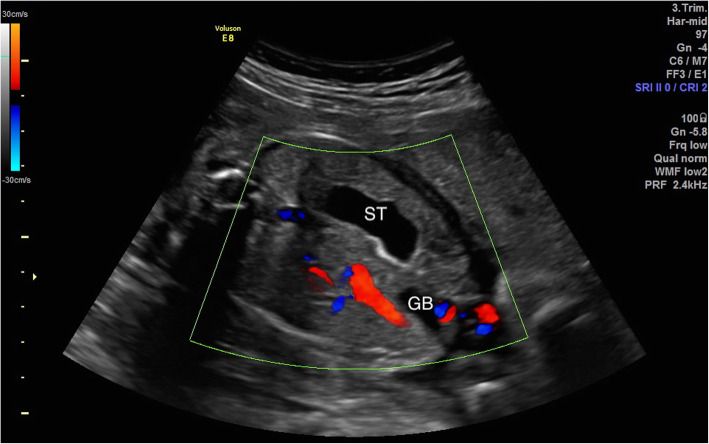
Persistent right umbilical vein (PRUV) is characterized by atresia of the left umbilical vein while the right umbilical vein remains open. Given the limited sample size of most studies, the incidence of PRUV and the status of concomitant anomalies may not be fully reflected. Thus, we studied the incidence of fetal PRUV and its concomitant anomalies on a larger scale using our hospital database. This study hoped to address the following questions: Does PRUV increase the risk of fetal anomalies? If the PRUV fetus also has a single umbilical artery (SUA), does the risk of fetal anomaly increase further? What is the positive predictive value of PRUV for fetal anomalies?
This retrospective study analyzed 756 cases of fetal PRUV at our hospital from January 2007 to April 2017. Prenatal ultrasound and color Doppler images were assessed. All PRUV fetuses underwent echocardiography and detailed ultrasound examinations of other systems. Newborn status was obtained via the database or by telephone follow-up.
A total of 435,428 pregnant women underwent prenatal ultrasonography at 16-40 weeks, the incidence of fetal PRUV was 0.17%, and 102 fetuses (13.5%) developed other anomalies. Two complicated cases had trisomy 18. PRUV was associated with a higher incidence of fetal anomalies. When fetal anomalies were classified by body systems, PRUV was associated with a higher incidence of cardiovascular, nervous, urinary, skeletal, digestive, and respiratory system anomalies. The positive predictive values of a PRUV for any fetal anomalies and cardiovascular anomalies were 13.5% (95%CI, 11.2-16.2%) and 5.4% (95%CI, 4.0-7.3%), respectively. SUA further increases the risk of PRUV fetuses with other anomalies and cardiovascular anomalies.
Detailed prenatal ultrasonography and echocardiography should be performed in fetuses with PRUV to rule out anomalies in other systems. When the PRUV is combined with SUA, echocardiography is particularly important. Fetuses with complicated PRUV should undergo chromosomal examination. Although isolated fetal PRUV prognosis is good, complicated PRUV prognosis depends on the type and severity of the concomitant anomalies.
持续性右脐静脉(PRUV)的特征为左脐静脉闭塞,而右脐静脉仍保持开放。由于大多数研究的样本量有限,PRUV 的发生率以及合并异常的情况可能无法得到充分反映。因此,我们利用医院数据库,对胎儿 PRUV 及其合并异常的发生率进行了更大规模的研究。本研究旨在回答以下问题:PRUV 是否会增加胎儿畸形的风险?如果 PRUV 胎儿还存在单脐动脉(SUA),胎儿畸形的风险是否会进一步增加?PRUV 对胎儿畸形的阳性预测值是多少?
本回顾性研究分析了 2007 年 1 月至 2017 年 4 月我院 756 例胎儿 PRUV 的病例资料。评估产前超声及彩色多普勒图像。所有 PRUV 胎儿均行超声心动图及其他系统的详细超声检查。通过数据库或电话随访获取新生儿情况。
共有 435428 名孕妇在 16-40 周行产前超声检查,胎儿 PRUV 的发生率为 0.17%,102 例(13.5%)胎儿发生其他异常。2 例复杂病例存在 18 三体。PRUV 与胎儿畸形的发生率较高有关。当按身体系统对胎儿畸形进行分类时,PRUV 与心血管、神经、泌尿、骨骼、消化和呼吸系统畸形的发生率较高有关。PRUV 对任何胎儿畸形和心血管畸形的阳性预测值分别为 13.5%(95%CI,11.2-16.2%)和 5.4%(95%CI,4.0-7.3%)。SUA 进一步增加了 PRUV 胎儿合并其他畸形和心血管畸形的风险。
对于 PRUV 胎儿应行详细的产前超声和超声心动图检查,以排除其他系统的异常。当 PRUV 合并 SUA 时,超声心动图尤为重要。复杂 PRUV 胎儿应行染色体检查。虽然孤立性胎儿 PRUV 预后良好,但复杂性 PRUV 的预后取决于合并异常的类型和严重程度。