Division of Gynecologic Oncology, Department of Obstetrics and Gynecology, Stanford University School of Medicine, 300 Pasteur Drive, H302, Stanford, CA, 94305, USA.
BMC Res Notes. 2020 Oct 12;13(1):480. doi: 10.1186/s13104-020-05314-9.
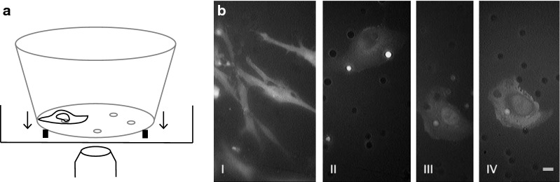
Cancer cell metastasis determines disease prognosis. During cancer cell metastasis, the cancer cell and the cancer cell nucleus have to undergo extreme shape changes. To monitor shape changes of cancer cells and cancer cell nuclei and the positioning of the cancer cell nucleus during cancer cell invasion, a customized invasion assay with 8-μm pores and reconstituted basal membrane was imaged using fluorescence live-cell microscopy.
The observed cells changed their shape from a distinct fibroblast-like spindle shape to an amoeboid shape without polarization immediately after the passage through an 8-μm pore of the invasion assay. During the process of invasion, the cancer cell centered the cancer cell nucleus over the 8-μm pore, and cancer cell nucleus and adjacent cytoplasmic areas moved first through such a pore. Seemingly testing if the largest and least deformable organelle may fit, the cancer cell nucleus led the way through the porous membrane of the invasion assay.
癌细胞转移决定着疾病的预后。在癌细胞转移过程中,癌细胞及其细胞核必须经历极端的形状变化。为了监测癌细胞和癌细胞核的形状变化以及癌细胞核在癌细胞侵袭过程中的定位,使用带有 8μm 孔的定制侵袭测定法和重建的基底膜,通过荧光活细胞显微镜进行成像。
观察到的细胞在穿过侵袭测定法的 8μm 孔后,立即从明显的成纤维细胞样纺锤形变为非极化的阿米巴样形状。在侵袭过程中,癌细胞将癌细胞核定位于 8μm 孔的中心,然后癌细胞核和相邻的细胞质区域首先通过该孔移动。癌细胞核似乎在测试最大和最不易变形的细胞器是否可以通过,从而引领着穿过侵袭测定法的多孔膜。