National-Regional Key Technology Engineering Laboratory for Medical Ultrasound, School of Biomedical Engineering, Health Science Center, Shenzhen University, Shenzhen, 518060, China.
Department of Biomedical Engineering and Environmental Sciences, National Tsing Hua University, Hsinchu, 30013 Taiwan.
Theranostics. 2020 Sep 26;10(25):11794-11819. doi: 10.7150/thno.44152. eCollection 2020.
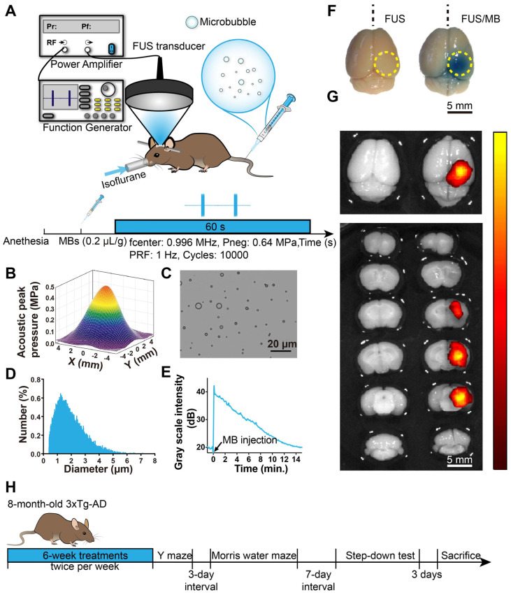
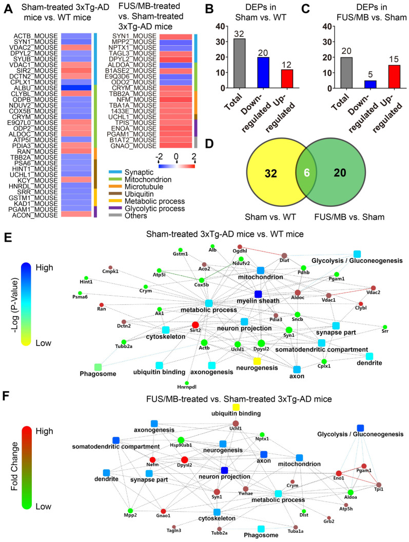
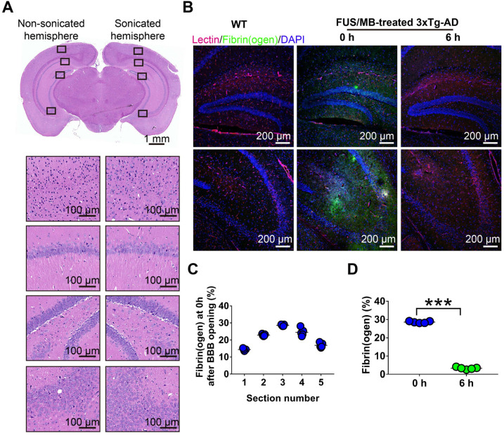
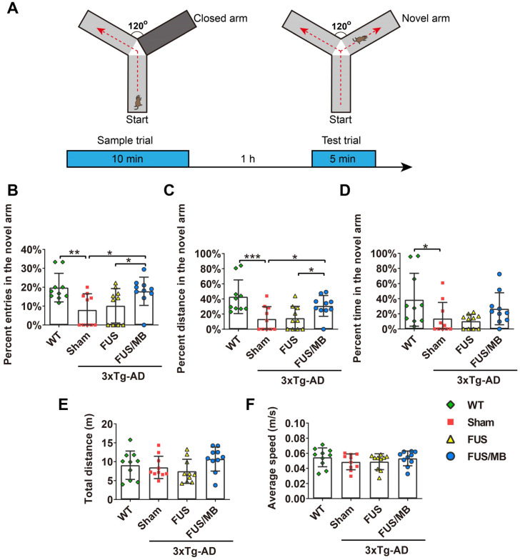
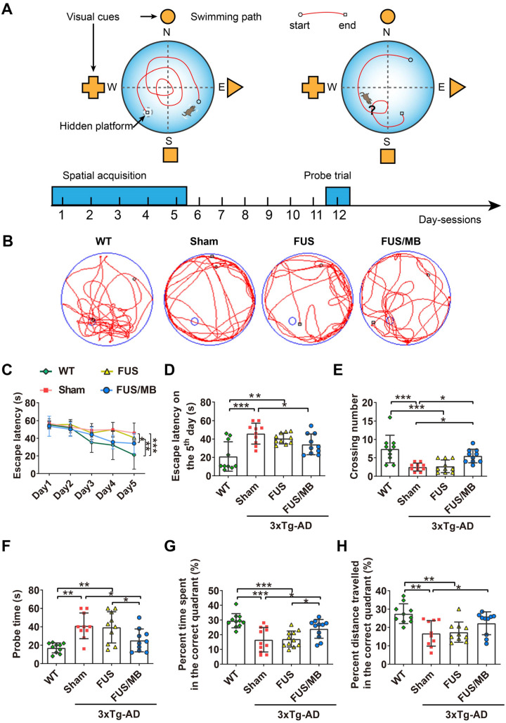
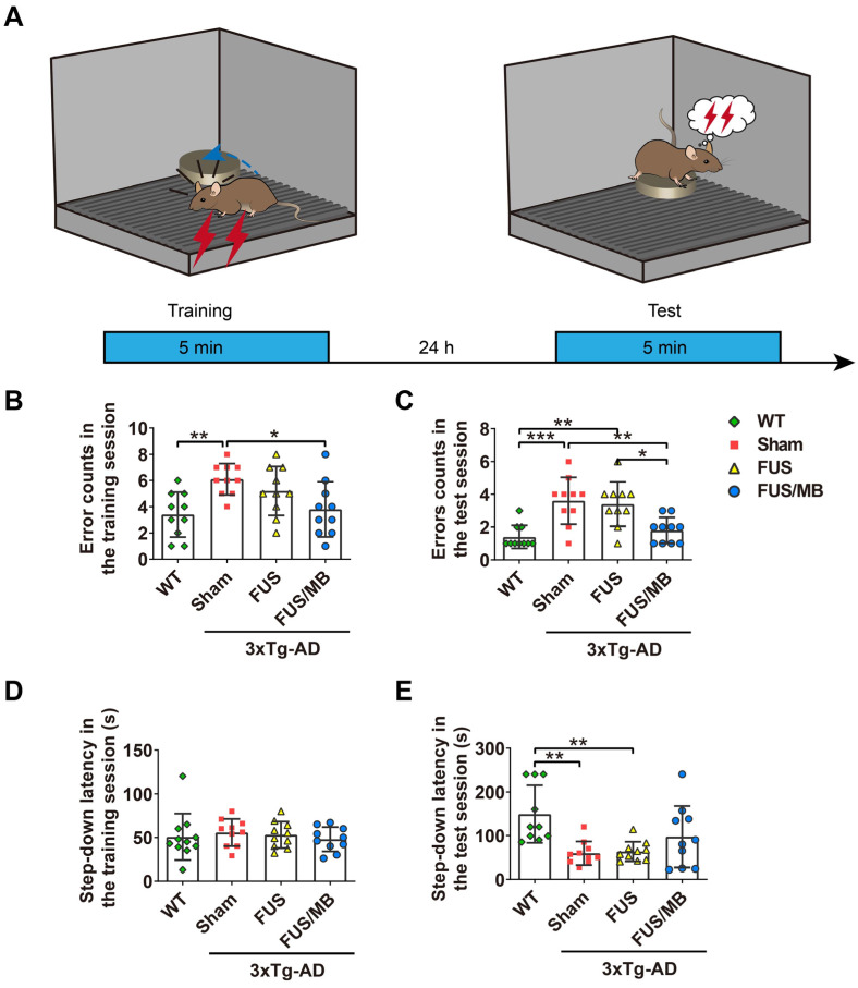
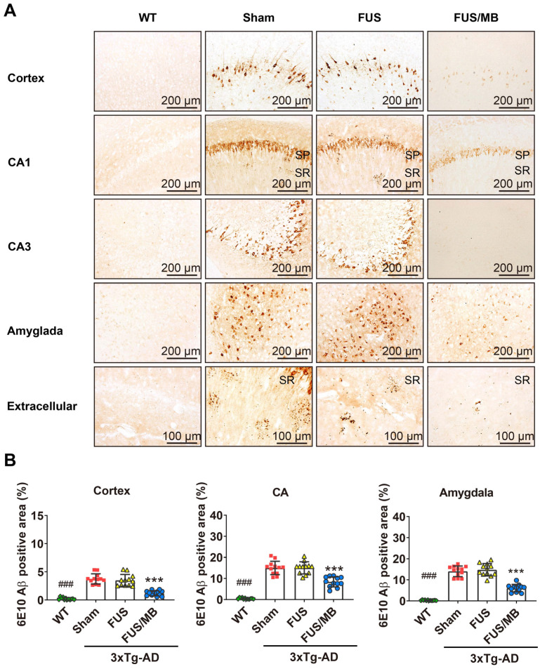
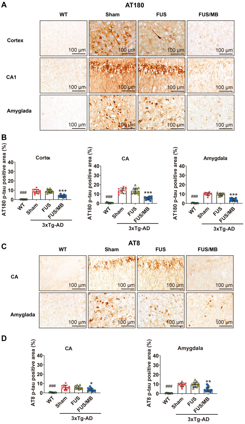
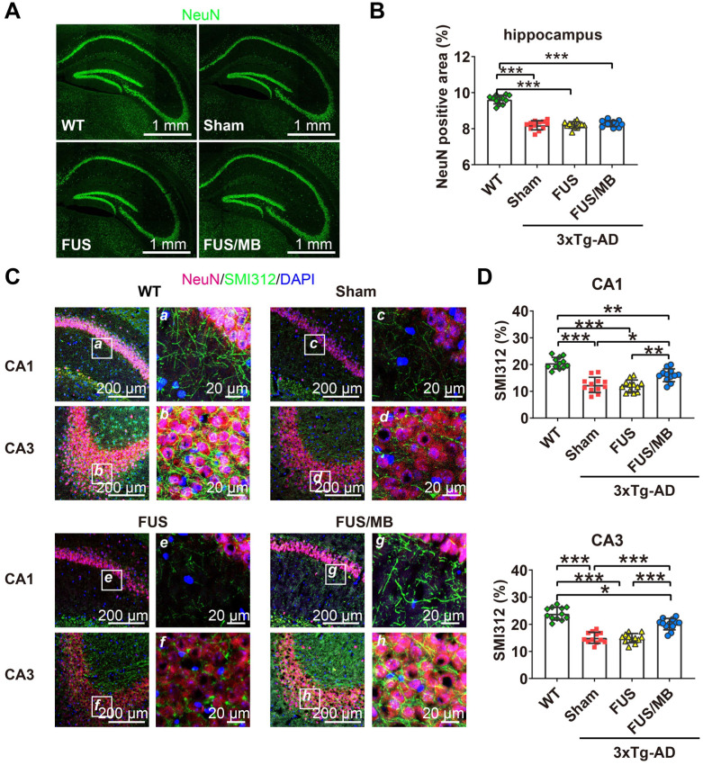
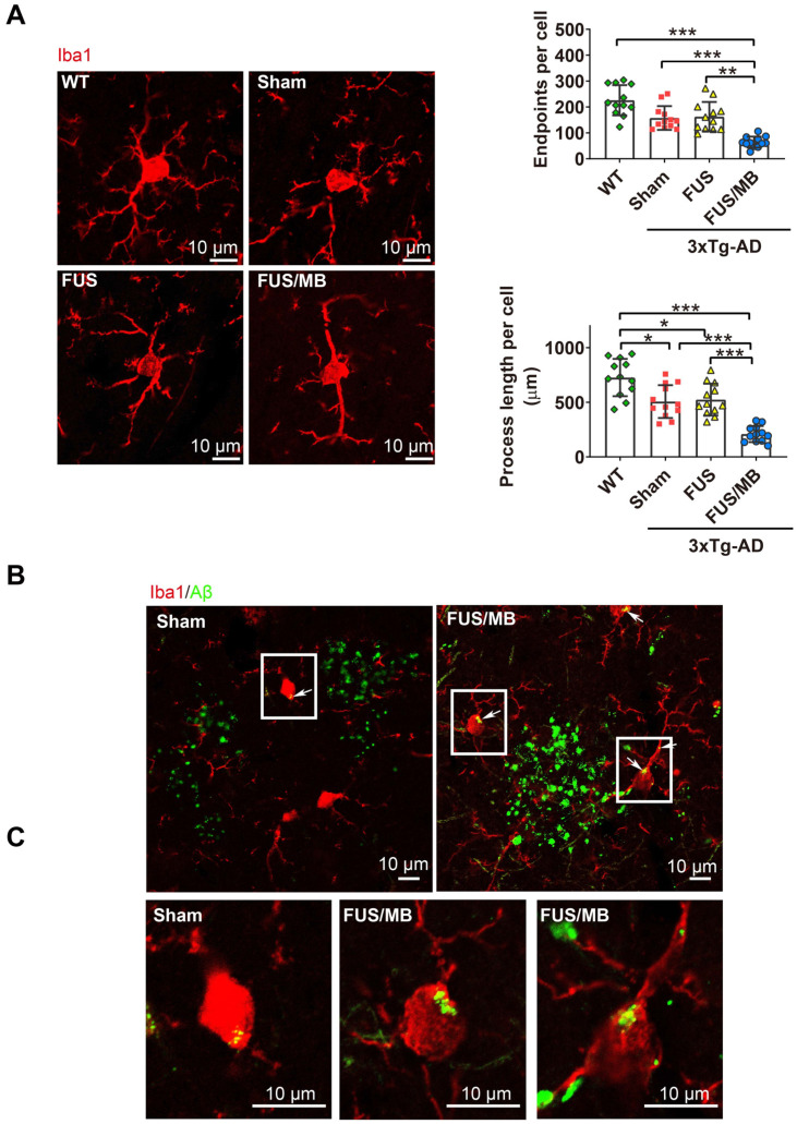
Alzheimer's disease (AD) is a progressive neurodegenerative disease manifested by cognitive impairment. As a unique approach to open the blood-brain barrier (BBB) noninvasively and temporarily, a growing number of studies showed that low-intensity focused ultrasound in combination with microbubbles (FUS/MB), in the absence of therapeutic agents, is capable of ameliorating amyloid or tau pathology, concurrent with improving memory deficits of AD animal models. However, the effects of FUS/MB on both the two pathologies simultaneously, as well as the memory behaviors, have not been reported so far. In this study, female triple transgenic AD (3×Tg-AD) mice at eight months of age with both amyloid-β (Aβ) deposits and tau phosphorylation were treated by repeated FUS/MB in the unilateral hippocampus twice per week for six weeks. The memory behaviors were investigated by the Y maze, the Morris water maze and the step-down passive avoidance test following repeated FUS/MB treatments. Afterwards, the involvement of Aβ and tau pathology were assessed by immunohistochemical analysis. Neuronal health and phagocytosis of Aβ deposits by microglia in the hippocampus were examined by confocal microscopy. Further, hippocampal proteomic alterations were analyzed by employing two-dimensional fluorescence difference gel electrophoresis (2D-DIGE) combined with mass spectrometry. The three independent memory tasks were indicative of evident learning and memory impairments in eight-month-old 3×Tg-AD mice, which developed intraneuronal Aβ, extracellular diffuse Aβ deposits and phosphorylated tau in the hippocampus and amygdala. Following repeated FUS/MB treatments, significant improvement in learning and memory ability of the 3×Tg-AD mice was achieved. Amelioration in both Aβ deposits and phosphorylated tau in the sonicated hemisphere was induced in FUS/MB-treated 3×Tg-AD mice. Albeit without increase in neuron density, enhancement in axonal neurofilaments emerged from the FUS/MB treatment. Confocal microscopy revealed activated microglia engulfing Aβ deposits in the FUS/MB-treated hippocampus. Further, proteomic analysis revealed 20 differentially expressed proteins, associated with glycolysis, neuron projection, mitochondrial pathways, metabolic process and ubiquitin binding etc., in the hippocampus between FUS/MB-treated and sham-treated 3×Tg-AD mice. Our findings reinforce the positive therapeutic effects on AD models with both Aβ and tau pathology induced by FUS/MB-mediated BBB opening, further supporting the potential of this treatment regime for clinical applications.
阿尔茨海默病(AD)是一种以认知障碍为特征的进行性神经退行性疾病。作为一种独特的无创、暂态打开血脑屏障(BBB)的方法,越来越多的研究表明,低强度聚焦超声联合微泡(FUS/MB)在没有治疗剂的情况下,能够改善 AD 动物模型的淀粉样蛋白或 tau 病理学,同时改善记忆缺陷。然而,到目前为止,还没有报道 FUS/MB 对这两种病理学以及记忆行为的同时影响。在这项研究中,8 月龄的雌性三转基因 AD(3×Tg-AD)小鼠双侧海马接受重复 FUS/MB 治疗,每周两次,共 6 周。治疗后通过 Y 迷宫、Morris 水迷宫和跳下被动回避测试评估记忆行为。之后,通过免疫组织化学分析评估淀粉样蛋白和 tau 病理学的参与情况。通过共聚焦显微镜检查海马神经元健康状况和小胶质细胞吞噬淀粉样蛋白沉积情况。进一步通过二维荧光差异凝胶电泳(2D-DIGE)联合质谱分析检测海马蛋白质组学改变。 三个独立的记忆任务表明 8 月龄 3×Tg-AD 小鼠存在明显的学习和记忆障碍,这些小鼠在海马和杏仁核中出现了神经元内淀粉样蛋白、细胞外弥散淀粉样蛋白沉积和磷酸化 tau。经过重复 FUS/MB 治疗,3×Tg-AD 小鼠的学习和记忆能力得到显著改善。在 FUS/MB 治疗的 3×Tg-AD 小鼠中,超声处理半球的淀粉样蛋白沉积和磷酸化 tau 均得到改善。尽管神经元密度没有增加,但 FUS/MB 治疗后轴突神经丝增强。共聚焦显微镜显示 FUS/MB 治疗的海马中活化的小胶质细胞吞噬淀粉样蛋白沉积。此外,蛋白质组学分析显示,在 FUS/MB 治疗和假手术处理的 3×Tg-AD 小鼠的海马中,有 20 种差异表达蛋白与糖酵解、神经元投射、线粒体途径、代谢过程和泛素结合等有关。 我们的研究结果强化了 FUS/MB 介导的 BBB 开放对 AD 模型中同时存在淀粉样蛋白和 tau 病理学的积极治疗效果,进一步支持了该治疗方案在临床应用中的潜力。