Institute of Medical Science, University of Toronto, Toronto, Ontario M5S 1A8, Canada.
Department of Neurology, Beth Israel Deaconess Medical Center and Division of Sleep Medicine, Harvard Medical School, Boston, MA 02215.
eNeuro. 2020 Oct 14;7(6). doi: 10.1523/ENEURO.0451-19.2020. Print 2020 Nov-Dec.
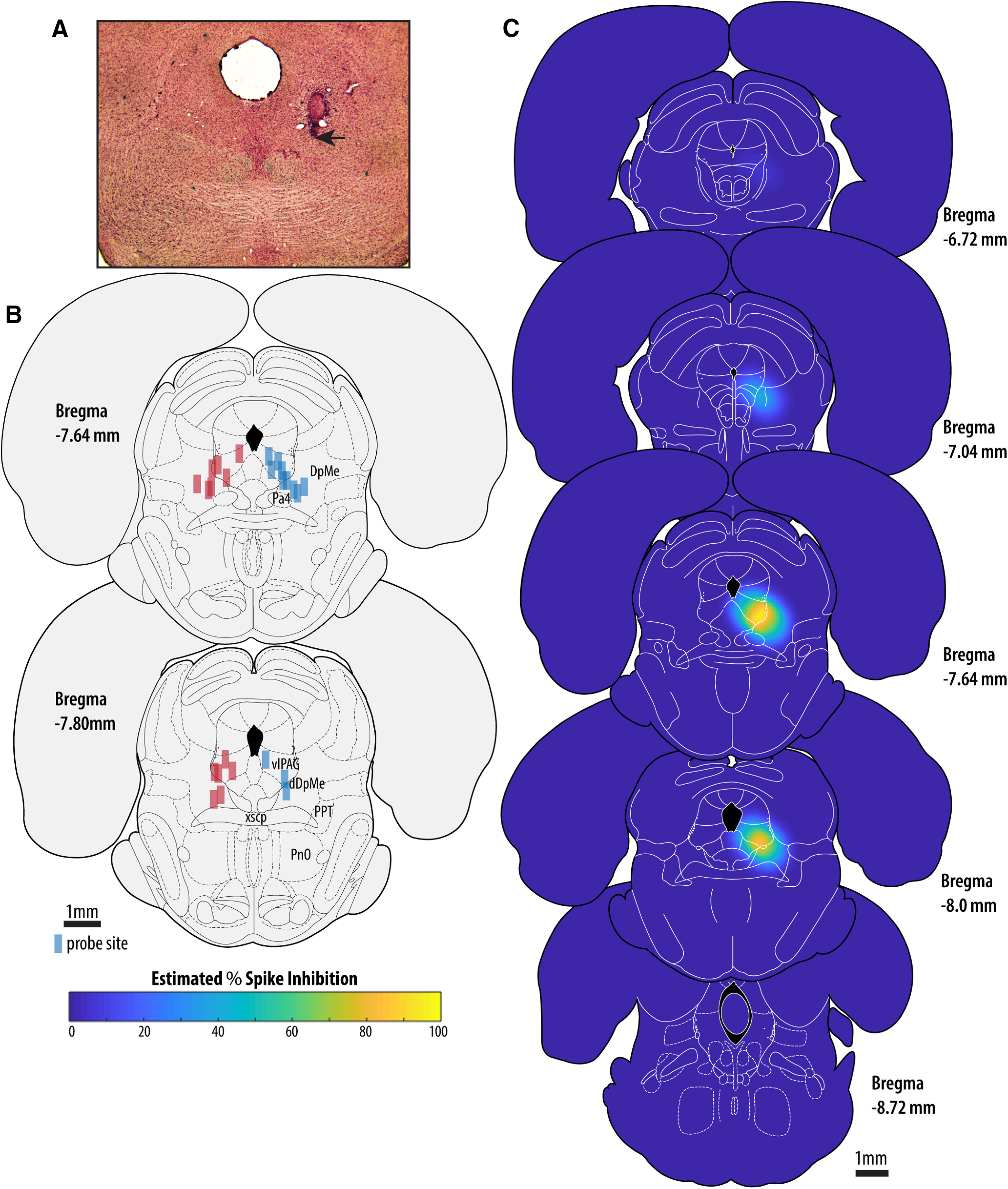
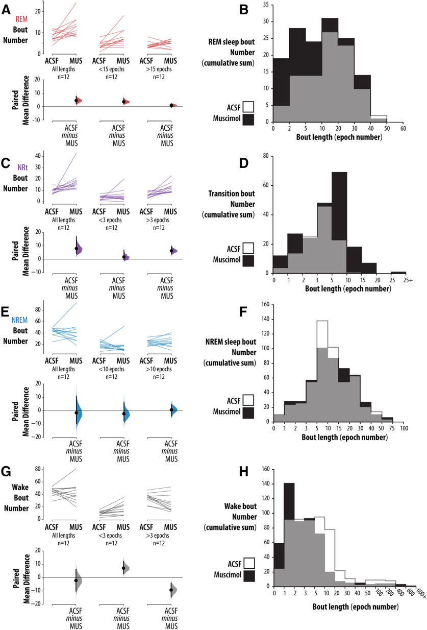
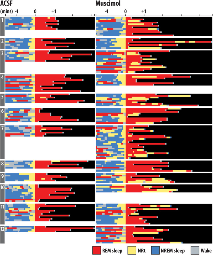
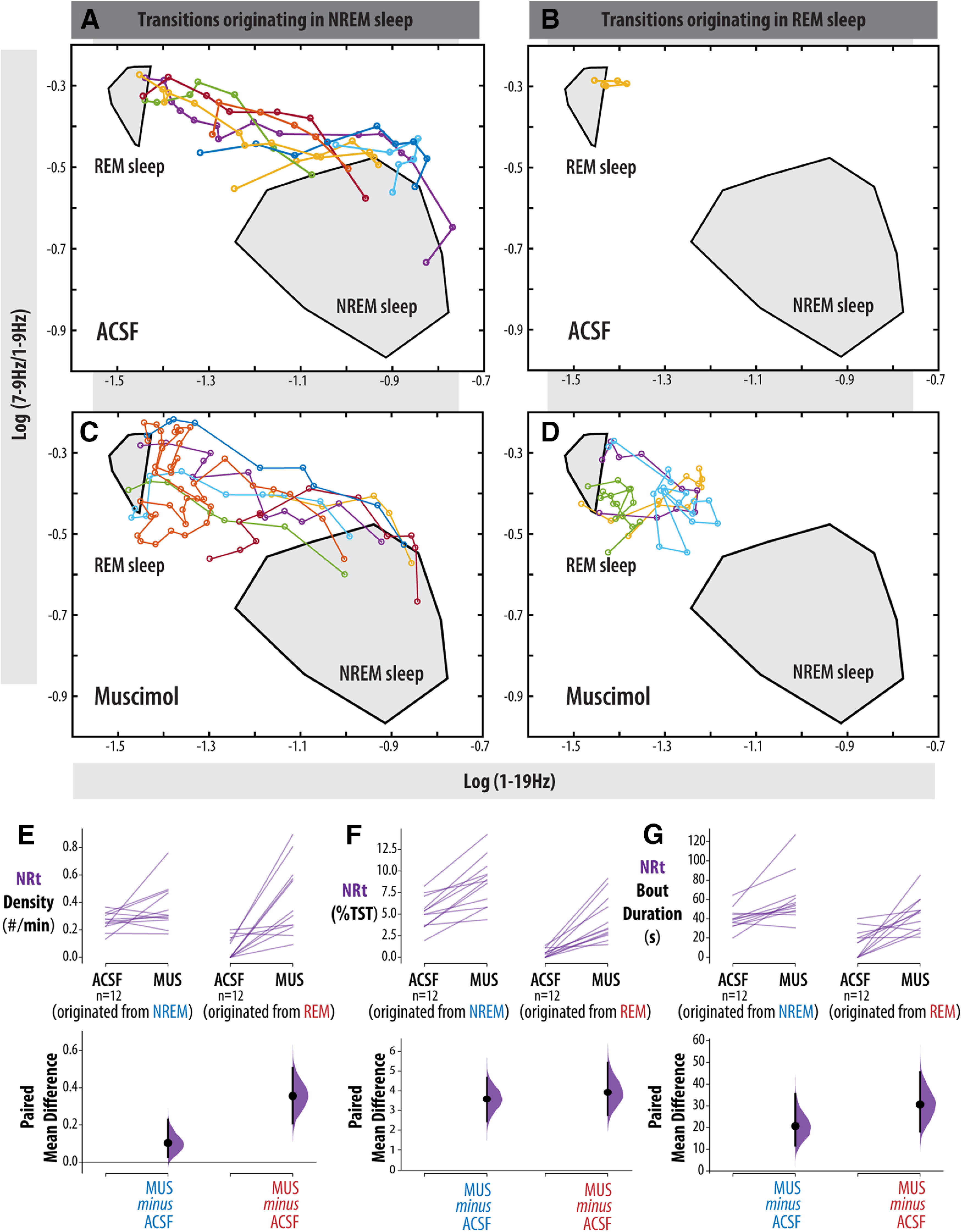
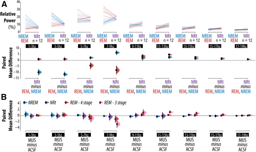
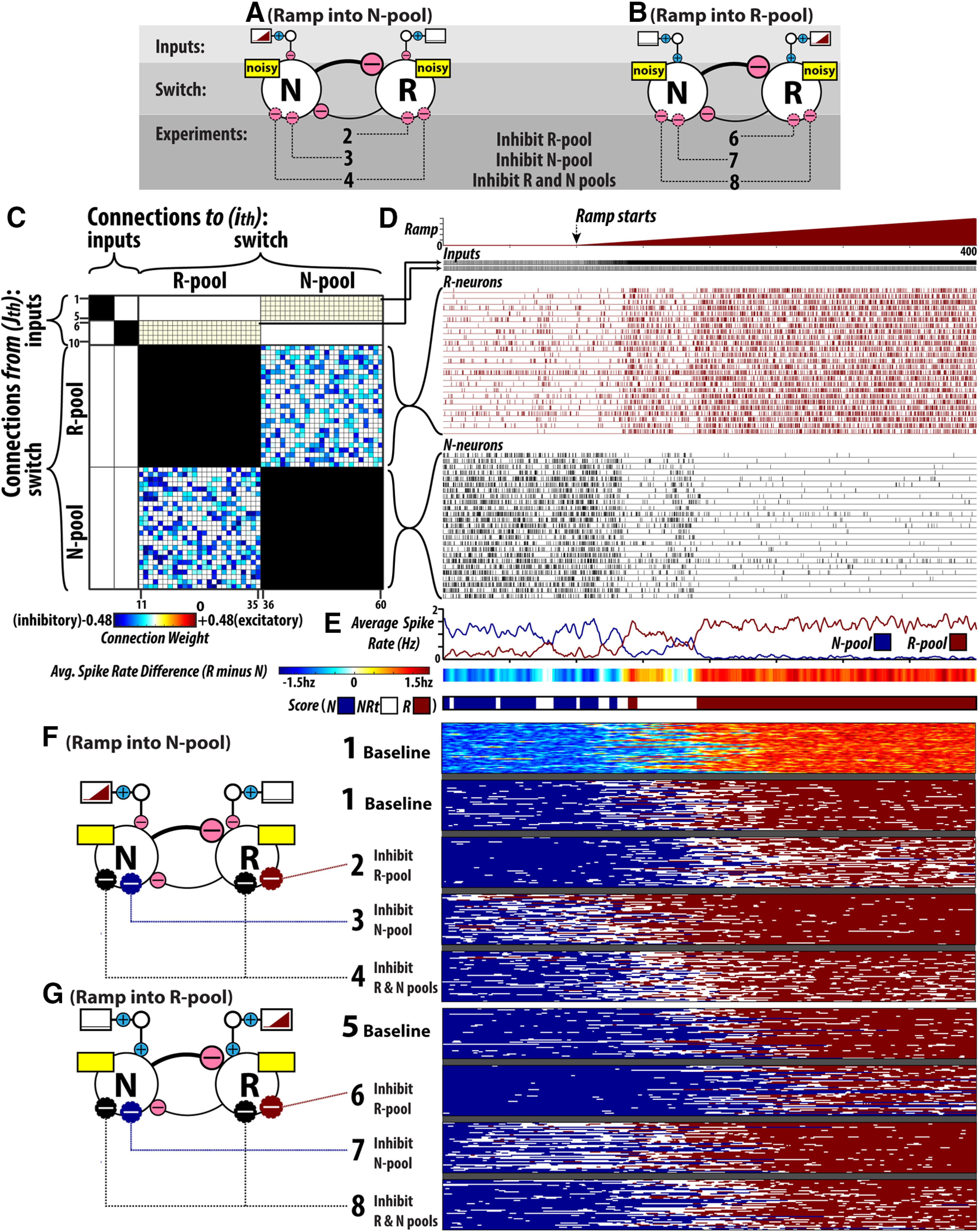
Neurons of the ventrolateral periaqueductal gray (vlPAG) and adjacent deep mesencephalic reticular nucleus (DpMe) are implicated in the control of sleep-wake state and are hypothesized components of a flip-flop circuit that maintains sleep bistability by preventing the overexpression of non-rapid eye movement (NREM)/REM sleep intermediary states (NRt). To determine the contribution of vlPAG/DpMe neurons in maintaining sleep bistability we combined computer simulations of flip-flop circuitry with focal inactivation of vlPAG/DpMe neurons by microdialysis delivery of the GABA receptor agonist muscimol in freely behaving male rats ( = 25) instrumented for electroencephalographic and electromyographic recording. REM sleep was enhanced by muscimol at the vlPAG/DpMe, consistent with previous studies; however, our analyses of NRt dynamics and those produced by flop-flop circuit simulations show that current thinking is too narrowly focused on the contribution of REM sleep-inactive populations toward vlPAG/DpMe involvement in REM sleep control. We found that much of the muscimol-mediated increase in REM sleep was more appropriately classified as NRt. This loss of sleep bistability was accompanied by fragmentation of REM sleep, as evidenced by an increased number of short REM sleep bouts. REM sleep fragmentation stemmed from an increased number and duration of NRt bouts originating in REM sleep. By contrast, NREM sleep bouts were not likewise fragmented by vlPAG/DpMe inactivation. In flip-flop circuit simulations, these changes could not be replicated through inhibition of the REM sleep-inactive population alone. Instead, combined suppression of REM sleep active and inactive vlPAG/DpMe subpopulations was required to replicate the changes in NRt dynamics.
腹外侧导水管周围灰质(vlPAG)和相邻的深部脑网状核(DpMe)神经元参与睡眠-觉醒状态的控制,并被假设为维持睡眠双稳定性的翻转电路的组成部分,通过防止非快速眼动(NREM)/快速眼动(REM)睡眠中间状态(NRt)的过度表达来维持睡眠双稳定性。为了确定 vlPAG/DpMe 神经元在维持睡眠双稳定性中的贡献,我们将翻转电路的计算机模拟与通过微透析向自由行为的雄性大鼠(n=25)的 vlPAG/DpMe 神经元施加 GABA 受体激动剂 muscimol 的局部失活相结合。REM 睡眠在 vlPAG/DpMe 处被 muscimol 增强,这与以前的研究一致;然而,我们对 NRt 动力学的分析以及翻转电路模拟产生的分析表明,目前的思维过于狭隘地集中在 REM 睡眠不活跃群体对 vlPAG/DpMe 参与 REM 睡眠控制的贡献上。我们发现,大部分由 muscimol 介导的 REM 睡眠增加更适合归类为 NRt。这种睡眠双稳定性的丧失伴随着 REM 睡眠的碎片化,这表现在 REM 睡眠短插曲的数量增加。REM 睡眠碎片化源于 REM 睡眠中起源的 NRt 插曲的数量和持续时间增加。相比之下,vlPAG/DpMe 失活不会使 NREM 睡眠插曲同样碎片化。在翻转电路模拟中,通过单独抑制 REM 睡眠不活跃群体,无法复制这些变化。相反,需要抑制 REM 睡眠活跃和不活跃的 vlPAG/DpMe 亚群的联合抑制来复制 NRt 动力学的变化。