Department of Radiation Therapy, National Cancer Centre, Singapore.
Department of Medical Imaging and Radiation Sciences, Monash University, Clayton, Australia.
J Med Radiat Sci. 2021 Jun;68(2):203-210. doi: 10.1002/jmrs.442. Epub 2020 Oct 15.
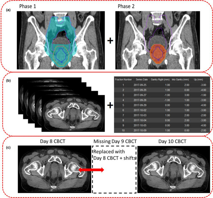
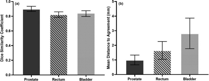
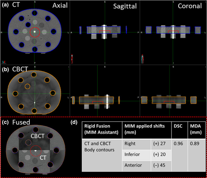
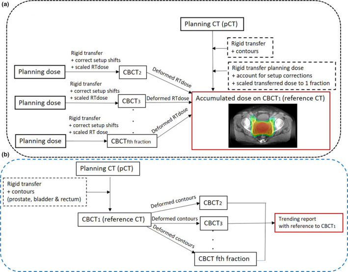
An automated dose accumulation and contour propagation workflow using daily cone beam computed tomography (CBCTs) images for prostate cases that require pelvic lymph nodes irradiation (PLNs) was developed. This workflow was constructed using MIM® software with the intention to provide accurate dose transformations for plans with two different isocentres, whereby two sequential treatment phases were prescribed. The pre-processing steps for data extractions from treatment plans, CBCTs, determination of couch shift information and management of missing CBCTs are described. To ensure that the imported translational couch shifts were in the correct orientation and readable in MIM, phantom commissioning was performed. For dose transformation, rigid registration with corrected setup shifts and scaled fractional dose was performed for pCT to daily CBCTs, which were then deformed onto CBCT . Fractional dose summation resulted in the final accumulated dose for the patient allowing differences in dosimetry between the planned and accumulated dose to be analysed. Contour propagations of the prostate, bladder and rectum were performed within the same workflow. Transformed contours were then deformed onto daily CBCTs to generate trending reports for analysis, including Dice Similarity Coefficient (DSC) and Mean Distance to Agreement (MDA). Results obtained from phantom commissioning (DSC = 0.96, MDA = 0.89 mm) and geometrical analysis of the propagated contours for twenty patients; prostate (DSC: 0.9 ± 0.0, MDA: 1.0 ± 0.3 mm), rectum (DSC: 0.8 ± 0.1, mm, MDA: 1.7 ± 0.6 mm) and bladder (DSC: 0.8 ± 0.1, MDA: 2.8 ± 1.0 mm) were within clinically accepted tolerances for both DSC (>0.8) and MDA (< 0.3 mm). The developed workflow is being performed on a larger patient cohort for predictive model building, with the goal of correlating observed toxicity with the actual accumulated dose received by the patient.
开发了一种使用需要盆腔淋巴结照射(PLN)的前列腺病例的每日锥形束 CT(CBCT)图像进行自动剂量积累和轮廓传播的工作流程。该工作流程使用 MIM®软件构建,旨在为具有两个不同等中心的计划提供准确的剂量转换,其中规定了两个连续的治疗阶段。描述了从治疗计划、CBCT 中提取数据、确定治疗床移位信息和管理缺失 CBCT 的预处理步骤。为了确保导入的平移治疗床移位在 MIM 中处于正确的方向并且可读,进行了体模验证。为了进行剂量转换,对 pCT 到每日 CBCT 进行了带校正设置移位的刚性配准和缩放分数剂量,然后将其变形到 CBCT 上。分数剂量求和导致患者的最终累积剂量,从而可以分析计划剂量和累积剂量之间的剂量差异。在同一工作流程中进行了前列腺、膀胱和直肠的轮廓传播。然后将转换后的轮廓变形到每日 CBCT 上,以生成用于分析的趋势报告,包括 Dice 相似系数(DSC)和平均差异一致度(MDA)。从体模验证(DSC=0.96,MDA=0.89mm)和 20 名患者传播轮廓的几何分析中获得的结果;前列腺(DSC:0.9±0.0,MDA:1.0±0.3mm)、直肠(DSC:0.8±0.1,MDA:1.7±0.6mm)和膀胱(DSC:0.8±0.1,MDA:2.8±1.0mm)均在 DSC(>0.8)和 MDA(<0.3mm)的临床可接受容差范围内。正在对更大的患者队列执行开发的工作流程,以进行预测模型构建,目标是将观察到的毒性与患者实际接收到的累积剂量相关联。