• 文献检索
  • 文档翻译
  • 深度研究
  • 学术资讯
  • Suppr Zotero 插件Zotero 插件
  • 邀请有礼
  • 套餐&价格
  • 历史记录
应用&插件
Suppr Zotero 插件Zotero 插件浏览器插件Mac 客户端Windows 客户端微信小程序
定价
高级版会员购买积分包购买API积分包
服务
文献检索文档翻译深度研究API 文档MCP 服务
关于我们
关于 Suppr公司介绍联系我们用户协议隐私条款
关注我们

Suppr 超能文献

核心技术专利:CN118964589B侵权必究
粤ICP备2023148730 号-1Suppr @ 2026

文献检索

告别复杂PubMed语法,用中文像聊天一样搜索,搜遍4000万医学文献。AI智能推荐,让科研检索更轻松。

立即免费搜索

文件翻译

保留排版,准确专业,支持PDF/Word/PPT等文件格式,支持 12+语言互译。

免费翻译文档

深度研究

AI帮你快速写综述,25分钟生成高质量综述,智能提取关键信息,辅助科研写作。

立即免费体验

基于超声图像纹理与形态特征高效融合的乳腺良恶性肿瘤分类方法。

A Benign and Malignant Breast Tumor Classification Method via Efficiently Combining Texture and Morphological Features on Ultrasound Images.

机构信息

College of Engineering, Huaqiao University, Quanzhou 362021, China.

School of Medicine, Huaqiao University, Quanzhou 362021, China.

出版信息

Comput Math Methods Med. 2020 Oct 1;2020:5894010. doi: 10.1155/2020/5894010. eCollection 2020.

DOI:10.1155/2020/5894010
PMID:33062038
原文链接:https://pmc.ncbi.nlm.nih.gov/articles/PMC7547332/
Abstract

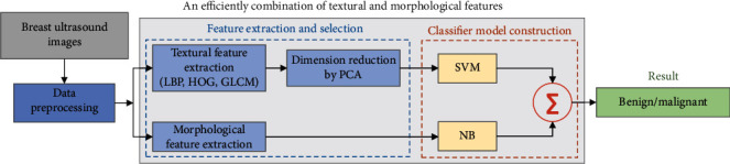
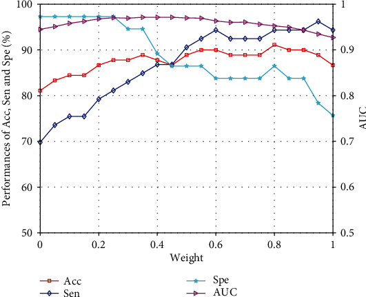
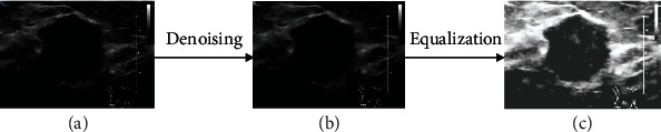
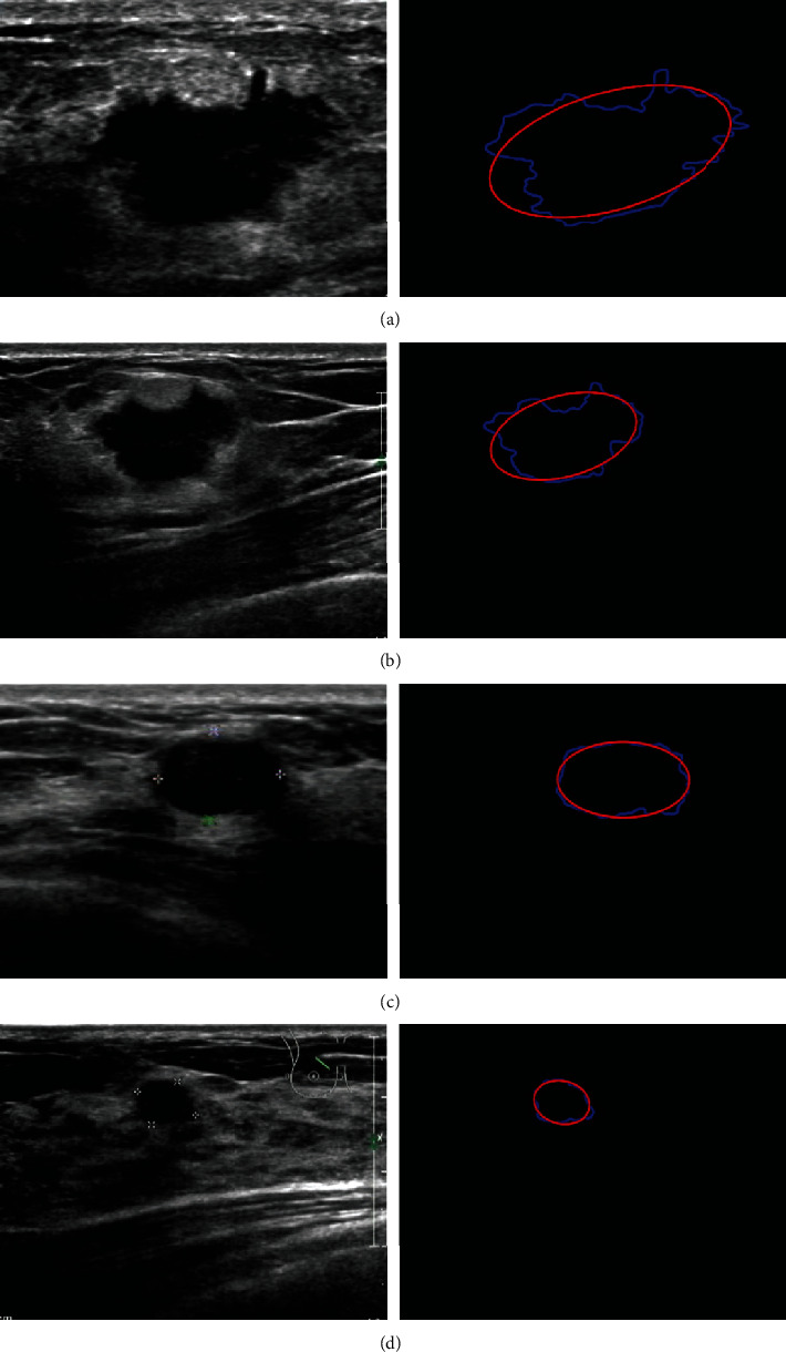
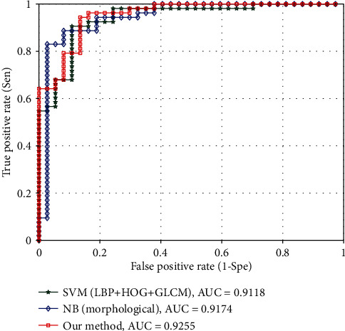
The classification of benign and malignant based on ultrasound images is of great value because breast cancer is an enormous threat to women's health worldwide. Although both texture and morphological features are crucial representations of ultrasound breast tumor images, their straightforward combination brings little effect for improving the classification of benign and malignant since high-dimensional texture features are too aggressive so that drown out the effect of low-dimensional morphological features. For that, an efficient texture and morphological feature combing method is proposed to improve the classification of benign and malignant. Firstly, both texture (i.e., (LBP), (HOG), and (GLCM)) and morphological (i.e., shape complexities) features of breast ultrasound images are extracted. Secondly, a (SVM) classifier working on texture features is trained, and a (NB) classifier acting on morphological features is designed, in order to exert the discriminative power of texture features and morphological features, respectively. Thirdly, the classification scores of the two classifiers (i.e., SVM and NB) are weighted fused to obtain the final classification result. The low-dimensional nonparameterized NB classifier is effectively control the parameter complexity of the entire classification system combine with the high-dimensional parametric SVM classifier. Consequently, texture and morphological features are efficiently combined. Comprehensive experimental analyses are presented, and the proposed method obtains a 91.11% accuracy, a 94.34% sensitivity, and an 86.49% specificity, which outperforms many related benign and malignant breast tumor classification methods.

摘要

基于超声图像的良性和恶性分类具有重要价值,因为乳腺癌是全世界女性健康的巨大威胁。尽管纹理和形态特征都是超声乳腺肿瘤图像的重要表示,但由于高维纹理特征过于激进,会淹没低维形态特征的效果,因此直接组合这两种特征对提高良性和恶性分类的效果不大。为此,提出了一种有效的纹理和形态特征组合方法来提高良性和恶性分类的效果。首先,提取乳腺超声图像的纹理(即 LBP、HOG 和 GLCM)和形态(即形状复杂度)特征。其次,训练基于纹理特征的支持向量机(SVM)分类器,并设计基于形态特征的朴素贝叶斯(NB)分类器,分别发挥纹理特征和形态特征的判别能力。然后,对两个分类器(SVM 和 NB)的分类得分进行加权融合,得到最终的分类结果。低维非参数化的 NB 分类器与高维参数化的 SVM 分类器相结合,可以有效地控制整个分类系统的参数复杂度。因此,可以有效地组合纹理和形态特征。进行了全面的实验分析,所提出的方法获得了 91.11%的准确率、94.34%的灵敏度和 86.49%的特异性,优于许多相关的良性和恶性乳腺肿瘤分类方法。

https://cdn.ncbi.nlm.nih.gov/pmc/blobs/2a95/7547332/0e112a7e6115/CMMM2020-5894010.007.jpg
https://cdn.ncbi.nlm.nih.gov/pmc/blobs/2a95/7547332/1bca624548e5/CMMM2020-5894010.001.jpg
https://cdn.ncbi.nlm.nih.gov/pmc/blobs/2a95/7547332/2b255449a5bd/CMMM2020-5894010.002.jpg
https://cdn.ncbi.nlm.nih.gov/pmc/blobs/2a95/7547332/698f5950f84c/CMMM2020-5894010.003.jpg
https://cdn.ncbi.nlm.nih.gov/pmc/blobs/2a95/7547332/a05aa272fa6c/CMMM2020-5894010.004.jpg
https://cdn.ncbi.nlm.nih.gov/pmc/blobs/2a95/7547332/2c9c340d535d/CMMM2020-5894010.005.jpg
https://cdn.ncbi.nlm.nih.gov/pmc/blobs/2a95/7547332/72315def3ae2/CMMM2020-5894010.006.jpg
https://cdn.ncbi.nlm.nih.gov/pmc/blobs/2a95/7547332/0e112a7e6115/CMMM2020-5894010.007.jpg

相似文献

1
A Benign and Malignant Breast Tumor Classification Method via Efficiently Combining Texture and Morphological Features on Ultrasound Images.基于超声图像纹理与形态特征高效融合的乳腺良恶性肿瘤分类方法。
Comput Math Methods Med. 2020 Oct 1;2020:5894010. doi: 10.1155/2020/5894010. eCollection 2020.
2
Robust phase-based texture descriptor for classification of breast ultrasound images.用于乳腺超声图像分类的基于稳健相位的纹理描述符
Biomed Eng Online. 2015 Mar 24;14:26. doi: 10.1186/s12938-015-0022-8.
3
Differentiation of fat-poor angiomyolipoma from clear cell renal cell carcinoma in contrast-enhanced MDCT images using quantitative feature classification.基于定量特征分类的 MDCT 增强图像鉴别乏脂性血管平滑肌脂肪瘤与透明细胞肾细胞癌
Med Phys. 2017 Jul;44(7):3604-3614. doi: 10.1002/mp.12258. Epub 2017 Jun 9.
4
Non-invasive automated 3D thyroid lesion classification in ultrasound: a class of ThyroScan™ systems.超声下非侵入式自动化 3D 甲状腺病变分类:一类 ThyroScan™ 系统。
Ultrasonics. 2012 Apr;52(4):508-20. doi: 10.1016/j.ultras.2011.11.003. Epub 2011 Nov 25.
5
Computer-assisted lip diagnosis on Traditional Chinese Medicine using multi-class support vector machines.基于多类支持向量机的中医唇诊计算机辅助诊断。
BMC Complement Altern Med. 2012 Aug 16;12:127. doi: 10.1186/1472-6882-12-127.
6
Breast Tumor Classification Using Intratumoral Quantitative Ultrasound Descriptors.基于肿瘤内定量超声特征对乳腺肿瘤进行分类。
Comput Math Methods Med. 2022 Mar 7;2022:1633858. doi: 10.1155/2022/1633858. eCollection 2022.
7
Comparison of methods for texture analysis of QUS parametric images in the characterization of breast lesions.用于乳腺病变特征描述的定量超声(QUS)参数图像纹理分析方法的比较
PLoS One. 2020 Dec 31;15(12):e0244965. doi: 10.1371/journal.pone.0244965. eCollection 2020.
8
A comparison of methods for three-class mammograms classification.三类乳腺X光片分类方法的比较。
Technol Health Care. 2017 Aug 9;25(4):657-670. doi: 10.3233/THC-160805.
9
Computerized analysis of mammographic microcalcifications in morphological and texture feature spaces.乳腺钼靶微钙化在形态学和纹理特征空间中的计算机分析
Med Phys. 1998 Oct;25(10):2007-19. doi: 10.1118/1.598389.
10
SVM classifier of cervical histopathology images based on texture and morphological features.基于纹理和形态特征的宫颈组织病理学图像支持向量机分类器
Technol Health Care. 2023;31(1):69-80. doi: 10.3233/THC-220031.

引用本文的文献

1
Evaluation of the clinical efficacy of mammotome minimally invasive rotary cutting surgery in the treatment of benign breast tumors: a comparative study.麦默通微创旋切手术治疗乳腺良性肿瘤的临床疗效评估:一项对比研究
J Robot Surg. 2025 May 7;19(1):207. doi: 10.1007/s11701-025-02367-5.
2
AI's ongoing impact: Implications of AI's effects on health equity for women's healthcare providers.人工智能的持续影响:人工智能对女性医疗保健提供者的健康公平性影响的含义。
Rev Panam Salud Publica. 2025 Apr 9;49:e19. doi: 10.26633/RPSP.2025.19. eCollection 2025.
3
Classifying breast cancer and fibroadenoma tissue biopsies from paraffined stain-free slides by fractal biomarkers in Fourier Ptychographic Microscopy.

本文引用的文献

1
Effect of a Deep Learning Framework-Based Computer-Aided Diagnosis System on the Diagnostic Performance of Radiologists in Differentiating between Malignant and Benign Masses on Breast Ultrasonography.深度学习框架辅助诊断系统对乳腺超声良恶性肿块鉴别诊断中放射科医生诊断性能的影响。
Korean J Radiol. 2019 May;20(5):749-758. doi: 10.3348/kjr.2018.0530.
2
Automated diagnosis of breast ultrasonography images using deep neural networks.基于深度神经网络的乳腺超声图像自动诊断。
Med Image Anal. 2019 Feb;52:185-198. doi: 10.1016/j.media.2018.12.006. Epub 2018 Dec 20.
3
Global cancer statistics 2018: GLOBOCAN estimates of incidence and mortality worldwide for 36 cancers in 185 countries.
利用傅里叶叠层显微镜中的分形生物标志物对石蜡免染切片中的乳腺癌和纤维腺瘤组织活检进行分类。
Comput Struct Biotechnol J. 2024 Mar 24;24:225-236. doi: 10.1016/j.csbj.2024.03.019. eCollection 2024 Dec.
4
Radiomics to Differentiate Malignant and Benign Breast Lesions: A Systematic Review and Diagnostic Test Accuracy Meta-Analysis.基于影像组学鉴别乳腺良恶性病变:一项系统评价与诊断试验准确性的Meta分析
Cureus. 2023 Nov 18;15(11):e49015. doi: 10.7759/cureus.49015. eCollection 2023 Nov.
5
Benign and Malignant Breast Tumor Classification in Ultrasound and Mammography Images via Fusion of Deep Learning and Handcraft Features.通过深度学习与手工特征融合实现超声和乳腺X线摄影图像中乳腺良恶性肿瘤的分类
Entropy (Basel). 2023 Jun 28;25(7):991. doi: 10.3390/e25070991.
6
The Systematic Review of Artificial Intelligence Applications in Breast Cancer Diagnosis.人工智能在乳腺癌诊断中的应用系统评价
Diagnostics (Basel). 2022 Dec 23;13(1):45. doi: 10.3390/diagnostics13010045.
7
The diagnostic significance of circulating miRNAs and metabolite profiling in early prediction of breast cancer in Egyptian women.循环 miRNA 和代谢物特征分析在埃及女性乳腺癌早期预测中的诊断意义。
J Cancer Res Clin Oncol. 2023 Jul;149(8):5437-5451. doi: 10.1007/s00432-022-04492-2. Epub 2022 Dec 2.
8
An Effective Method for Lung Cancer Diagnosis from CT Scan Using Deep Learning-Based Support Vector Network.一种基于深度学习支持向量网络的利用CT扫描诊断肺癌的有效方法。
Cancers (Basel). 2022 Nov 6;14(21):5457. doi: 10.3390/cancers14215457.
9
Dual-Intended Deep Learning Model for Breast Cancer Diagnosis in Ultrasound Imaging.用于超声成像中乳腺癌诊断的双目标深度学习模型。
Cancers (Basel). 2022 May 27;14(11):2663. doi: 10.3390/cancers14112663.
10
Real-time denoising of ultrasound images based on deep learning.基于深度学习的超声图像实时去噪。
Med Biol Eng Comput. 2022 Aug;60(8):2229-2244. doi: 10.1007/s11517-022-02573-5. Epub 2022 Jun 7.
全球癌症统计数据 2018:GLOBOCAN 对全球 185 个国家/地区 36 种癌症的发病率和死亡率的估计。
CA Cancer J Clin. 2018 Nov;68(6):394-424. doi: 10.3322/caac.21492. Epub 2018 Sep 12.
4
A Radiomics Approach With CNN for Shear-Wave Elastography Breast Tumor Classification.基于卷积神经网络的剪切波弹性成像乳腺肿瘤分类的放射组学方法。
IEEE Trans Biomed Eng. 2018 Sep;65(9):1935-1942. doi: 10.1109/TBME.2018.2844188. Epub 2018 Jun 5.
5
Machine Learning in Ultrasound Computer-Aided Diagnostic Systems: A Survey.基于机器学习的超声计算机辅助诊断系统:综述
Biomed Res Int. 2018 Mar 4;2018:5137904. doi: 10.1155/2018/5137904. eCollection 2018.
6
Automated localization and segmentation techniques for B-mode ultrasound images: A review.B 型超声图像的自动定位和分割技术:综述。
Comput Biol Med. 2018 Jan 1;92:210-235. doi: 10.1016/j.compbiomed.2017.11.018. Epub 2017 Dec 6.
7
A computer-aided diagnosis system for breast ultrasound based on weighted BI-RADS classes.基于加权 BI-RADS 类别的乳腺超声计算机辅助诊断系统。
Comput Methods Programs Biomed. 2018 Jan;153:33-40. doi: 10.1016/j.cmpb.2017.10.004. Epub 2017 Oct 3.
8
Breast cancer statistics, 2017, racial disparity in mortality by state.乳腺癌统计数据,2017 年,按州划分的死亡率种族差异。
CA Cancer J Clin. 2017 Nov;67(6):439-448. doi: 10.3322/caac.21412. Epub 2017 Oct 3.
9
The adaptive computer-aided diagnosis system based on tumor sizes for the classification of breast tumors detected at screening ultrasound.基于肿瘤大小的自适应计算机辅助诊断系统,用于筛查超声检测出的乳腺肿瘤分类。
Ultrasonics. 2017 Apr;76:70-77. doi: 10.1016/j.ultras.2016.12.017. Epub 2016 Dec 28.
10
Fully Convolutional Networks for Semantic Segmentation.全卷积网络用于语义分割。
IEEE Trans Pattern Anal Mach Intell. 2017 Apr;39(4):640-651. doi: 10.1109/TPAMI.2016.2572683. Epub 2016 May 24.