• 文献检索
  • 文档翻译
  • 深度研究
  • 学术资讯
  • Suppr Zotero 插件Zotero 插件
  • 邀请有礼
  • 套餐&价格
  • 历史记录
应用&插件
Suppr Zotero 插件Zotero 插件浏览器插件Mac 客户端Windows 客户端微信小程序
定价
高级版会员购买积分包购买API积分包
服务
文献检索文档翻译深度研究API 文档MCP 服务
关于我们
关于 Suppr公司介绍联系我们用户协议隐私条款
关注我们

Suppr 超能文献

核心技术专利:CN118964589B侵权必究
粤ICP备2023148730 号-1Suppr @ 2026

文献检索

告别复杂PubMed语法,用中文像聊天一样搜索,搜遍4000万医学文献。AI智能推荐,让科研检索更轻松。

立即免费搜索

文件翻译

保留排版,准确专业,支持PDF/Word/PPT等文件格式,支持 12+语言互译。

免费翻译文档

深度研究

AI帮你快速写综述,25分钟生成高质量综述,智能提取关键信息,辅助科研写作。

立即免费体验

次级骨化中心诱导并保护生长板结构。

Secondary ossification center induces and protects growth plate structure.

机构信息

Department of Physiology and Pharmacology, Karolinska Institutet, Stockholm, Sweden.

Department of Evolutionary Morphology, Schmalhausen Institute of Zoology of NAS of Ukraine, Kiev, Ukraine.

出版信息

Elife. 2020 Oct 16;9:e55212. doi: 10.7554/eLife.55212.

DOI:10.7554/eLife.55212
PMID:33063669
原文链接:
https://pmc.ncbi.nlm.nih.gov/articles/PMC7581430/
Abstract

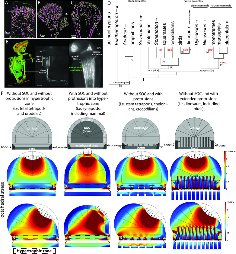
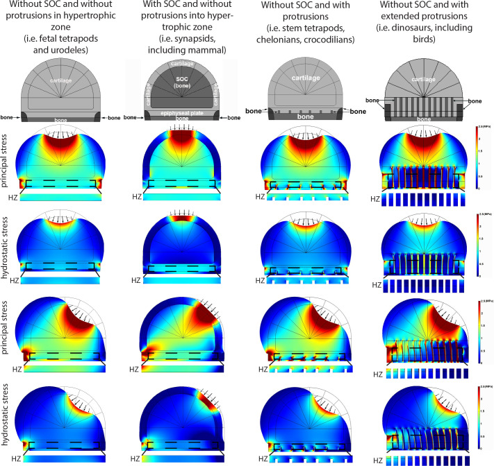
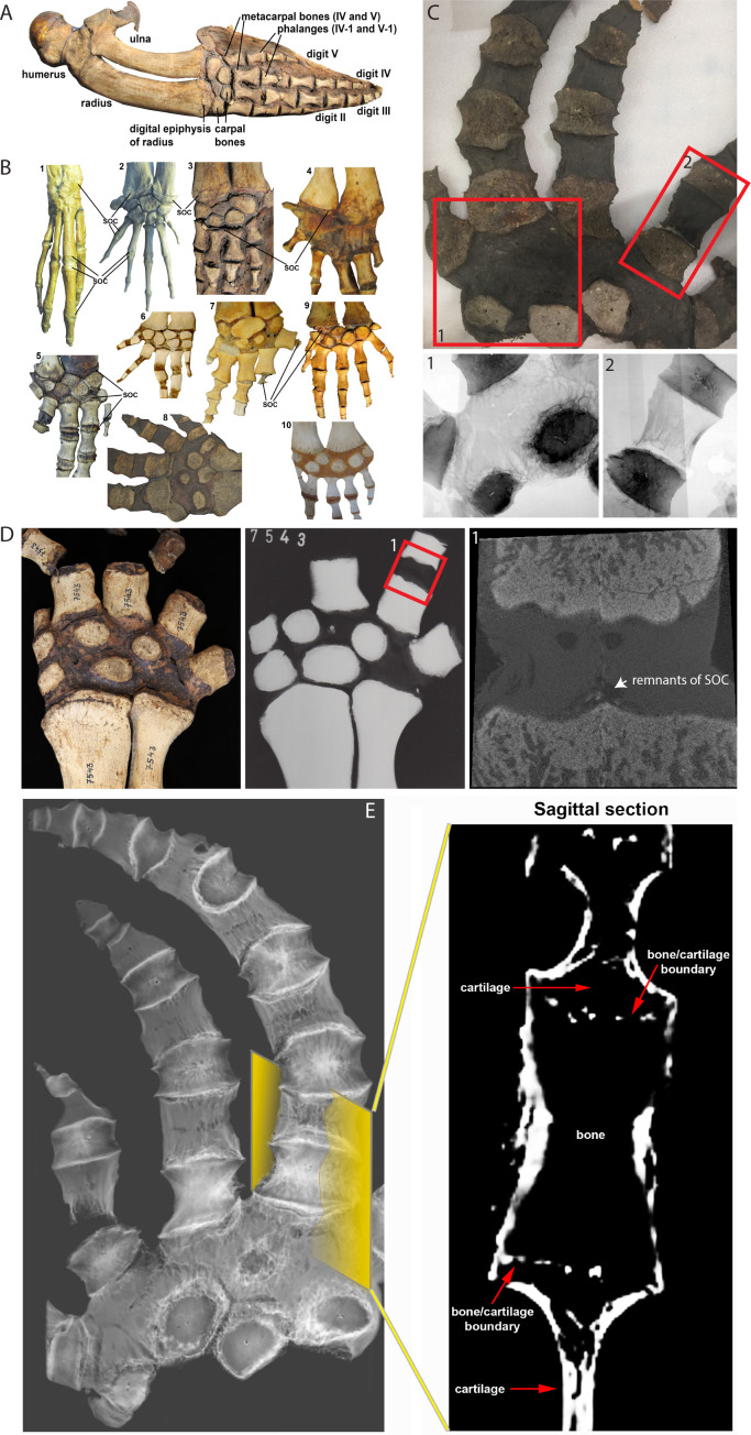
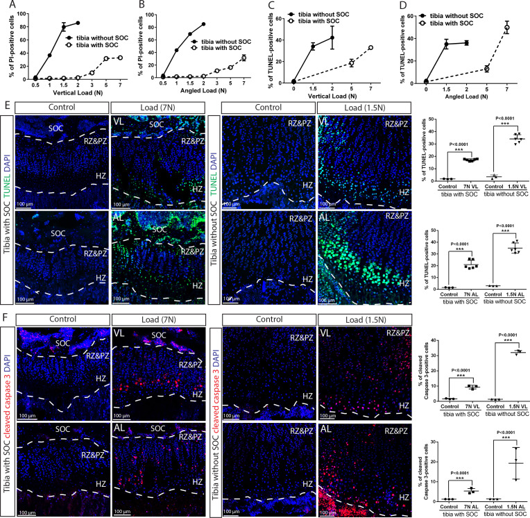
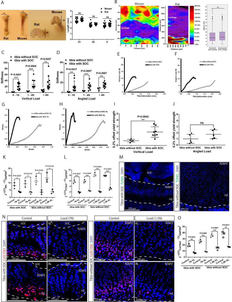
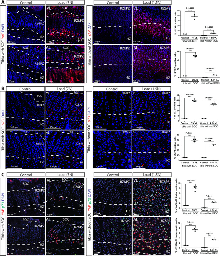
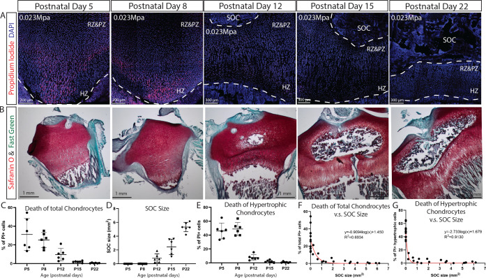
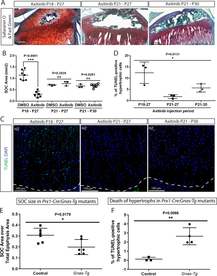
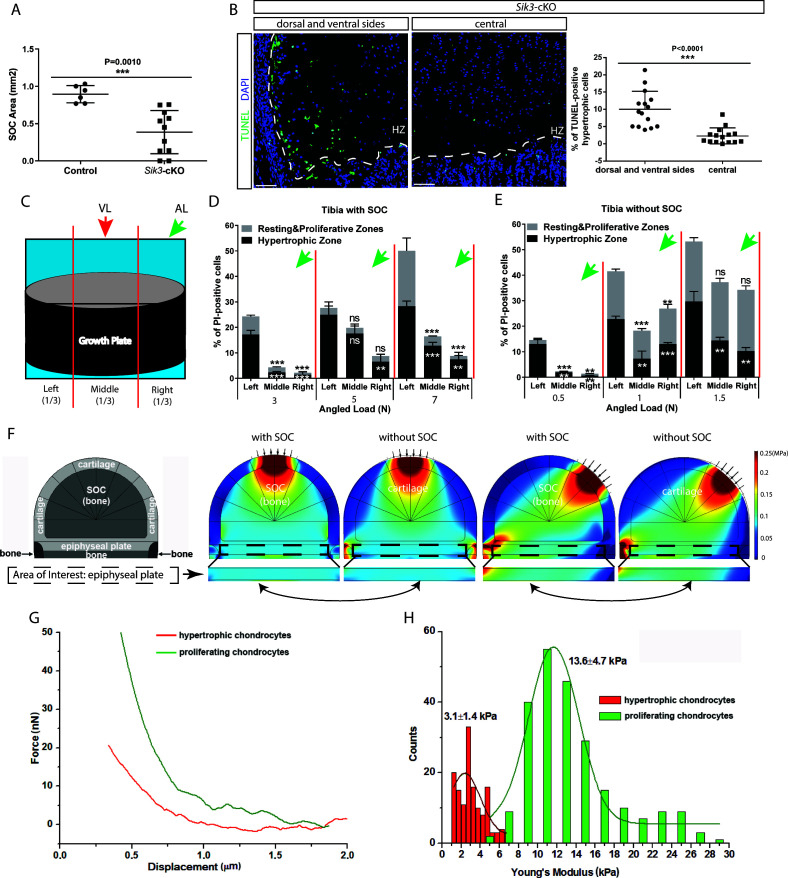
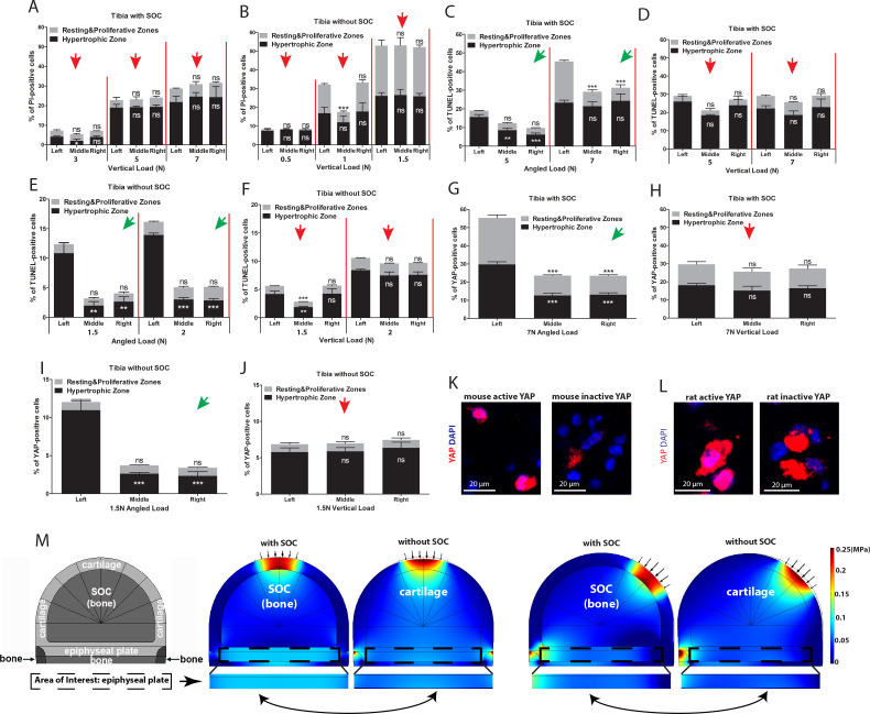
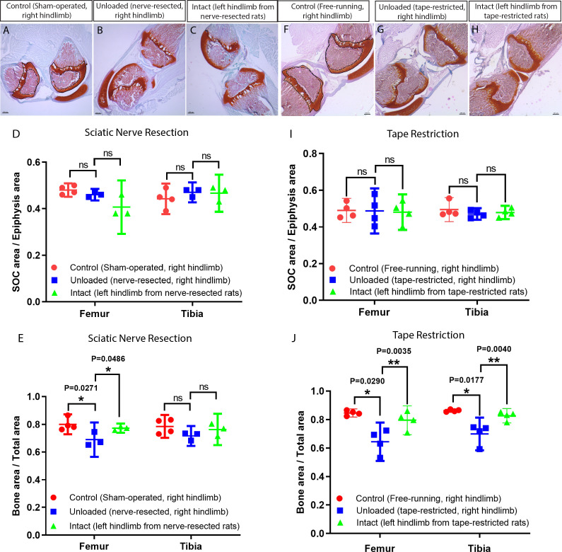
Growth plate and articular cartilage constitute a single anatomical entity early in development but later separate into two distinct structures by the secondary ossification center (SOC). The reason for such separation remains unknown. We found that evolutionarily SOC appears in animals conquering the land - amniotes. Analysis of the ossification pattern in mammals with specialized extremities (whales, bats, jerboa) revealed that SOC development correlates with the extent of mechanical loads. Mathematical modeling revealed that SOC reduces mechanical stress within the growth plate. Functional experiments revealed the high vulnerability of hypertrophic chondrocytes to mechanical stress and showed that SOC protects these cells from apoptosis caused by extensive loading. Atomic force microscopy showed that hypertrophic chondrocytes are the least mechanically stiff cells within the growth plate. Altogether, these findings suggest that SOC has evolved to protect the hypertrophic chondrocytes from the high mechanical stress encountered in the terrestrial environment.

摘要

生长板和关节软骨在发育早期构成一个单一的解剖实体,但后来通过次级骨化中心 (SOC) 分离成两个不同的结构。这种分离的原因尚不清楚。我们发现 SOC 出现在征服陆地的动物——羊膜动物中。对具有特殊四肢的哺乳动物(鲸鱼、蝙蝠、跳鼠)的骨化模式进行分析表明,SOC 的发展与机械负荷的程度有关。数学模型表明,SOC 降低了生长板内的机械应力。功能实验表明,肥大软骨细胞对机械应力非常敏感,并且 SOC 可以保护这些细胞免受过度负荷引起的细胞凋亡。原子力显微镜显示,肥大软骨细胞是生长板内机械硬度最低的细胞。总的来说,这些发现表明 SOC 的进化是为了保护肥大软骨细胞免受陆地环境中遇到的高机械应力的影响。

https://cdn.ncbi.nlm.nih.gov/pmc/blobs/50d0/7581430/0c3edcc86f2c/elife-55212-fig10-figsupp2.jpg
https://cdn.ncbi.nlm.nih.gov/pmc/blobs/50d0/7581430/43117c657123/elife-55212-fig1.jpg
https://cdn.ncbi.nlm.nih.gov/pmc/blobs/50d0/7581430/06591cb213a4/elife-55212-fig1-figsupp1.jpg
https://cdn.ncbi.nlm.nih.gov/pmc/blobs/50d0/7581430/421e54be0108/elife-55212-fig1-figsupp2.jpg
https://cdn.ncbi.nlm.nih.gov/pmc/blobs/50d0/7581430/2060773a34b5/elife-55212-fig2.jpg
https://cdn.ncbi.nlm.nih.gov/pmc/blobs/50d0/7581430/1983a29bd6b6/elife-55212-fig3.jpg
https://cdn.ncbi.nlm.nih.gov/pmc/blobs/50d0/7581430/54ce1a2eec3a/elife-55212-fig4.jpg
https://cdn.ncbi.nlm.nih.gov/pmc/blobs/50d0/7581430/e66e097daca1/elife-55212-fig5.jpg
https://cdn.ncbi.nlm.nih.gov/pmc/blobs/50d0/7581430/63b603ddad37/elife-55212-fig5-figsupp1.jpg
https://cdn.ncbi.nlm.nih.gov/pmc/blobs/50d0/7581430/0eeff016fec5/elife-55212-fig5-figsupp2.jpg
https://cdn.ncbi.nlm.nih.gov/pmc/blobs/50d0/7581430/123fed884837/elife-55212-fig5-figsupp3.jpg
https://cdn.ncbi.nlm.nih.gov/pmc/blobs/50d0/7581430/a0891887dfa8/elife-55212-fig6.jpg
https://cdn.ncbi.nlm.nih.gov/pmc/blobs/50d0/7581430/036f901d1630/elife-55212-fig6-figsupp1.jpg
https://cdn.ncbi.nlm.nih.gov/pmc/blobs/50d0/7581430/888d09b2da44/elife-55212-fig7.jpg
https://cdn.ncbi.nlm.nih.gov/pmc/blobs/50d0/7581430/b0e8273044bb/elife-55212-fig8.jpg
https://cdn.ncbi.nlm.nih.gov/pmc/blobs/50d0/7581430/a09293fd1015/elife-55212-fig9.jpg
https://cdn.ncbi.nlm.nih.gov/pmc/blobs/50d0/7581430/a8fe3be559d8/elife-55212-fig9-figsupp1.jpg
https://cdn.ncbi.nlm.nih.gov/pmc/blobs/50d0/7581430/1d2915d4a2e0/elife-55212-fig10.jpg
https://cdn.ncbi.nlm.nih.gov/pmc/blobs/50d0/7581430/9944124f8414/elife-55212-fig10-figsupp1.jpg
https://cdn.ncbi.nlm.nih.gov/pmc/blobs/50d0/7581430/0c3edcc86f2c/elife-55212-fig10-figsupp2.jpg

相似文献

1
Secondary ossification center induces and protects growth plate structure.次级骨化中心诱导并保护生长板结构。
Elife. 2020 Oct 16;9:e55212. doi: 10.7554/eLife.55212.
2
Modulation of endochondral development of the distal femoral condyle by mechanical loading.机械负荷对股骨远端髁软骨内发育的调节作用。
J Orthop Res. 2006 Feb;24(2):229-41. doi: 10.1002/jor.20024.
3
Periarticular Mesenchymal Progenitors Initiate and Contribute to Secondary Ossification Center Formation During Mouse Long Bone Development.关节周围间充质祖细胞在小鼠长骨发育过程中启动并有助于次生骨化中心的形成。
Stem Cells. 2019 May;37(5):677-689. doi: 10.1002/stem.2975. Epub 2019 Feb 6.
4
SOX9 is a major negative regulator of cartilage vascularization, bone marrow formation and endochondral ossification.SOX9 是软骨血管生成、骨髓形成和软骨内骨化的主要负调控因子。
Development. 2010 Mar;137(6):901-11. doi: 10.1242/dev.045203.
5
mir-374-5p, mir-379-5p, and mir-503-5p Regulate Proliferation and Hypertrophic Differentiation of Growth Plate Chondrocytes in Male Rats.miR-374-5p、miR-379-5p 和 miR-503-5p 调控雄性大鼠生长板软骨细胞的增殖和肥大分化。
Endocrinology. 2018 Mar 1;159(3):1469-1478. doi: 10.1210/en.2017-00780.
6
Novel effects of CCN3 that may direct the differentiation of chondrocytes.CCN3 的新作用可能指导软骨细胞的分化。
FEBS Lett. 2011 Oct 3;585(19):3033-40. doi: 10.1016/j.febslet.2011.08.024. Epub 2011 Aug 23.
7
Leptin differentially regulates endochondral ossification in tibial and vertebral epiphyseal plates.瘦素在胫骨和椎骨骺板的软骨内骨化中发挥差异化调节作用。
Cell Biol Int. 2018 Feb;42(2):169-179. doi: 10.1002/cbin.10882. Epub 2017 Nov 9.
8
Annexin VIII is differentially expressed by chondrocytes in the mammalian growth plate during endochondral ossification and in osteoarthritic cartilage.膜联蛋白VIII在哺乳动物生长板软骨内成骨过程中以及骨关节炎软骨中,由软骨细胞呈差异性表达。
J Bone Miner Res. 2002 Oct;17(10):1851-8. doi: 10.1359/jbmr.2002.17.10.1851.
9
Sorting of growth plate chondrocytes allows the isolation and characterization of cells of a defined differentiation status.生长板软骨细胞的分拣允许分离和鉴定具有特定分化状态的细胞。
J Bone Miner Res. 2010 Jun;25(6):1267-81. doi: 10.1002/jbmr.30.
10
Primary culture of rat growth plate chondrocytes: an in vitro model of growth plate histotype, matrix vesicle biogenesis and mineralization.大鼠生长板软骨细胞的原代培养:生长板组织类型、基质小泡生物发生及矿化的体外模型
Bone. 2004 Jun;34(6):961-70. doi: 10.1016/j.bone.2004.02.010.

引用本文的文献

1
Physical and chemical niche of human growth plate for polarized bone development.用于极化骨发育的人类生长板的物理和化学微环境
Nat Commun. 2025 Aug 8;16(1):7328. doi: 10.1038/s41467-025-62711-z.
2
Single-cell expression profiling of bat wing development.蝙蝠翅膀发育的单细胞表达谱分析。
Nat Commun. 2025 Jul 18;16(1):6612. doi: 10.1038/s41467-025-61944-2.
3
Modeling endochondral ossification: Effects of mechanical loading and bone shape.软骨内成骨建模:机械负荷与骨形态的影响

本文引用的文献

1
The tuatara genome reveals ancient features of amniote evolution.圆口蜥基因组揭示了羊膜动物进化的古老特征。
Nature. 2020 Aug;584(7821):403-409. doi: 10.1038/s41586-020-2561-9. Epub 2020 Aug 5.
2
A radical switch in clonality reveals a stem cell niche in the epiphyseal growth plate.克隆性的根本转变揭示了骺板中的干细胞龛位。
Nature. 2019 Mar;567(7747):234-238. doi: 10.1038/s41586-019-0989-6. Epub 2019 Feb 27.
3
Gain-of-function mutation of microRNA-140 in human skeletal dysplasia.人类骨骼发育不良中 microRNA-140 的功能获得性突变。
J Orthop. 2025 May 31;68:197-218. doi: 10.1016/j.jor.2025.05.034. eCollection 2025 Oct.
4
Exploring the impact of mechanical stimuli on growth plate morphology and trabecular adaptation: A finite element approach.探索机械刺激对生长板形态和小梁适应性的影响:一种有限元方法。
J Orthop. 2025 Apr 28;63:228-237. doi: 10.1016/j.jor.2025.04.014. eCollection 2025 May.
5
Dynamics of postnatal bone development and epiphyseal synostosis in the caprine autopod.山羊足部产后骨骼发育及骨骺融合的动态变化
Dev Dyn. 2025 May 13. doi: 10.1002/dvdy.70038.
6
Dynamics of postnatal bone development and epiphyseal synostosis in the caprine autopod.山羊自足骨产后骨骼发育及骨骺融合的动态变化
bioRxiv. 2024 Dec 26:2024.12.26.630423. doi: 10.1101/2024.12.26.630423.
7
Timing of resting zone parathyroid hormone-related protein expression affects maintenance of the growth plate during secondary ossification: a computational study.静止区甲状旁腺激素相关蛋白表达的时机对二次骨化过程中生长板的维持有影响:一项计算研究
Biomech Model Mechanobiol. 2025 Feb;24(1):125-137. doi: 10.1007/s10237-024-01899-3. Epub 2024 Nov 16.
8
Relaxed selection in evolution of genes regulating limb development gives clue to variation in forelimb morphology of cetaceans and other mammals.基因调控肢体发育的进化中的松弛选择为鲸类和其他哺乳动物前肢形态的变异提供了线索。
Proc Biol Sci. 2024 Oct;291(2032):20241106. doi: 10.1098/rspb.2024.1106. Epub 2024 Oct 9.
9
Endochondral Ossification for Spinal Fusion: A Novel Perspective from Biological Mechanisms to Clinical Applications.脊柱融合的软骨内成骨:从生物学机制到临床应用的新视角
J Pers Med. 2024 Sep 9;14(9):957. doi: 10.3390/jpm14090957.
10
Postcranial anatomy of (Reptilia: Ichthyosauria) from the Middle Triassic Besano Formation of Monte San Giorgio (Italy/Switzerland), with implications for reconstructing the swimming styles of Triassic ichthyosaurs.来自意大利/瑞士蒙特圣乔治中三叠世贝萨诺组的(爬行纲:鱼龙目)的颅后解剖结构,及其对重建三叠纪鱼龙游泳方式的意义。
Swiss J Palaeontol. 2024;143(1):32. doi: 10.1186/s13358-024-00330-9. Epub 2024 Sep 10.
Nat Med. 2019 Apr;25(4):583-590. doi: 10.1038/s41591-019-0353-2. Epub 2019 Feb 25.
4
Constitutive stimulatory G protein activity in limb mesenchyme impairs bone growth.肢体间质中组成性刺激 G 蛋白活性会损害骨骼生长。
Bone. 2018 May;110:230-237. doi: 10.1016/j.bone.2018.02.016. Epub 2018 Feb 20.
5
Life history of the stem tetrapod Acanthostega revealed by synchrotron microtomography.同步辐射显微断层扫描揭示的四足动物棘螈的生活史。
Nature. 2016 Sep 15;537(7620):408-411. doi: 10.1038/nature19354. Epub 2016 Sep 7.
6
Mapping the Nonreciprocal Micromechanics of Individual Cells and the Surrounding Matrix Within Living Tissues.绘制活组织内单个细胞与周围基质的非互易微力学图谱。
Sci Rep. 2016 Apr 12;6:24272. doi: 10.1038/srep24272.
7
Effect of growth plate geometry and growth direction on prediction of proximal femoral morphology.生长板几何形状和生长方向对股骨近端形态预测的影响。
J Biomech. 2016 Jun 14;49(9):1613-1619. doi: 10.1016/j.jbiomech.2016.03.039. Epub 2016 Mar 31.
8
Role of Thyroid Hormones in Skeletal Development and Bone Maintenance.甲状腺激素在骨骼发育和骨骼维持中的作用。
Endocr Rev. 2016 Apr;37(2):135-87. doi: 10.1210/er.2015-1106. Epub 2016 Feb 10.
9
A pathway to bone: signaling molecules and transcription factors involved in chondrocyte development and maturation.通往骨骼的途径:参与软骨细胞发育和成熟的信号分子及转录因子
Development. 2015 Mar 1;142(5):817-31. doi: 10.1242/dev.105536.
10
Hypertrophic chondrocytes can become osteoblasts and osteocytes in endochondral bone formation.肥大的软骨细胞可在软骨内骨形成中成为成骨细胞和骨细胞。
Proc Natl Acad Sci U S A. 2014 Aug 19;111(33):12097-102. doi: 10.1073/pnas.1302703111. Epub 2014 Aug 4.