Department of Biomedical and Biotechnological Sciences, School of Medicine, University of Catania, via S.Sofia 97, 95123 Catania, Italy.
Int J Mol Sci. 2020 Oct 13;21(20):7528. doi: 10.3390/ijms21207528.
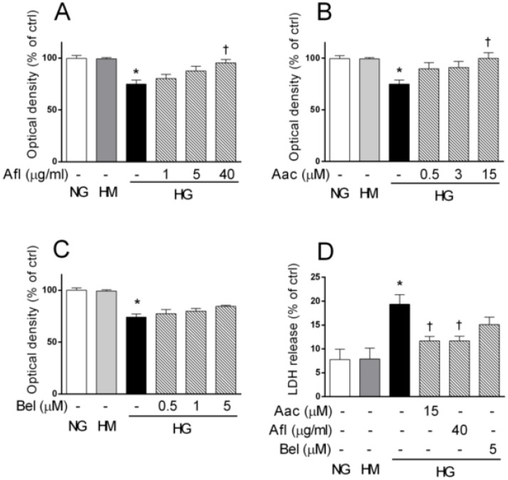
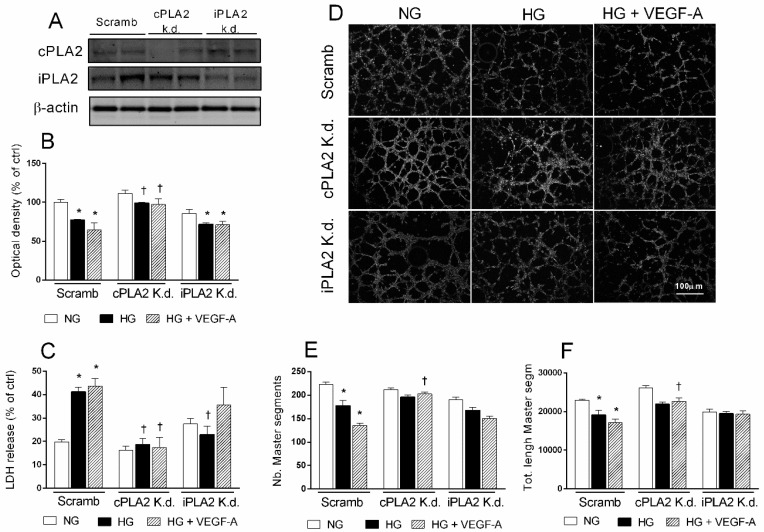
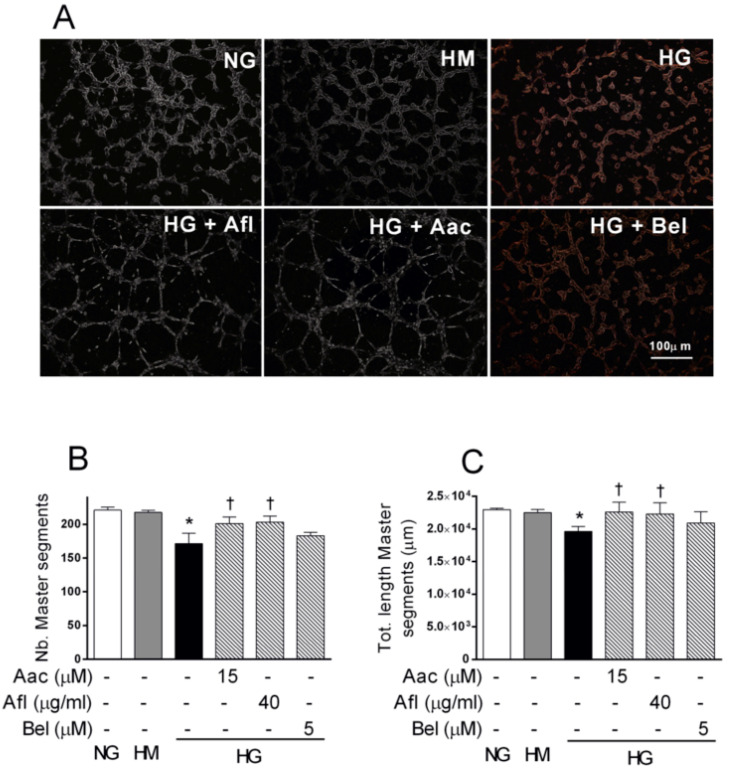
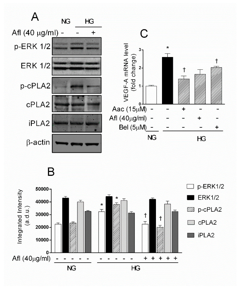
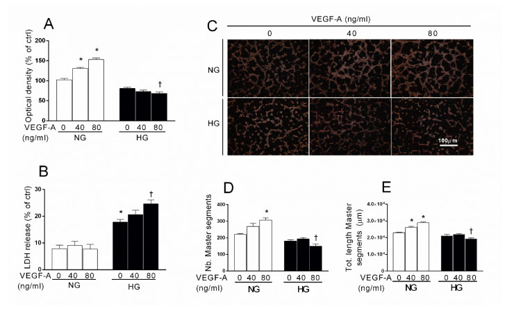
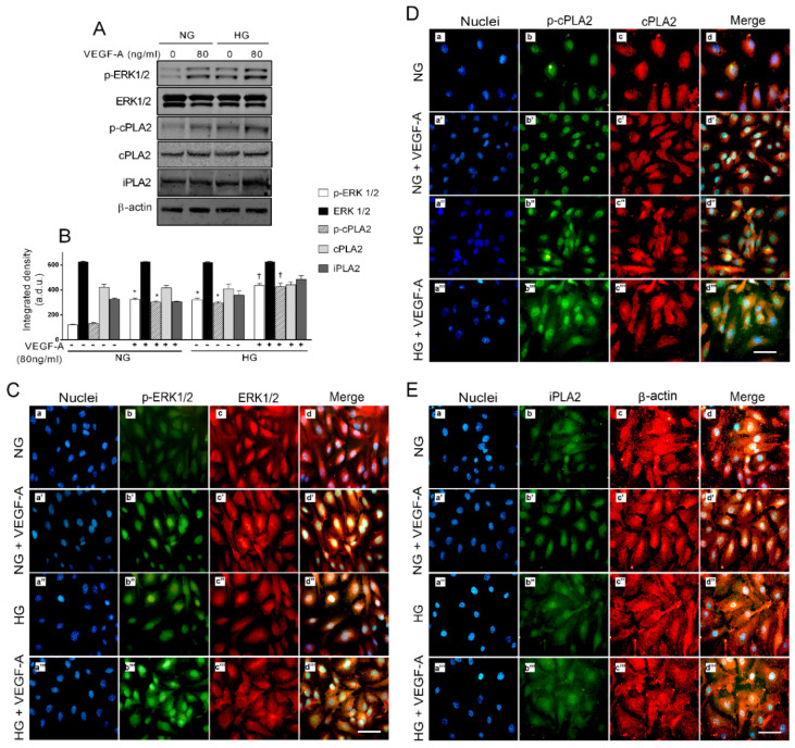
Early blood retinal barrier (BRB) dysfunction induced by hyperglycemia was related to increased pro-inflammatory activity of phospholipase A2 (PLA2) and the upregulation of vascular endothelial growth factor A (VEGF-A). Here, we tested the role of VEGF-A in high glucose (HG)-induced damage of human retinal endothelial cells (HRECs) mediated by Ca++-dependent (cPLA2) and Ca++-independent (iPLA2) PLA2s. HRECs were treated with normal glucose (5 mM, NG) or high glucose (25 mM, HG) for 48 h with or without the VEGF-trap Aflibercept (Afl, 40 µg/mL), the cPLA2 inhibitor arachidonoyl trifluoromethyl ketone (AACOCF3; 15 µM), the iPLA2 inhibitor bromoenol lactone (BEL; 5 µM), or VEGF-A (80 ng/mL). Both Afl and AACOCF3 prevented HG-induced damage (MTT and LDH release), impairment of angiogenic potential (tube-formation), and expression of VEGF-A mRNA. Furthermore, Afl counteracted HG-induced increase of phospho-ERK and phospho-cPLA2 (immunoblot). VEGF-A in HG-medium increased glucose toxicity, through upregulation of phospho-ERK, phospho-cPLA2, and iPLA2 (about 55%, 45%, and 50%, respectively); immunocytochemistry confirmed the activation of these proteins. cPLA2 knockdown by siRNA entirely prevented cell damage induced by HG or by HG plus VEGF-A, while iPLA2 knockdown produced a milder protective effect. These data indicate that VEGF-A mediates the early glucose-induced damage in retinal endothelium through the involvement of ERK1/2/PLA2 axis activation.
早期高血糖引起的血视网膜屏障(BRB)功能障碍与磷脂酶 A2(PLA2)的促炎活性增加和血管内皮生长因子 A(VEGF-A)的上调有关。在这里,我们测试了 VEGF-A 在高葡萄糖(HG)诱导的人视网膜内皮细胞(HRECs)损伤中的作用,该损伤由 Ca++依赖性(cPLA2)和 Ca++非依赖性(iPLA2)PLA2 介导。用正常葡萄糖(5 mM,NG)或高葡萄糖(25 mM,HG)处理 HRECs 48 h,用或不用 VEGF 陷阱 aflibercept(Afl,40 µg/mL)、cPLA2 抑制剂 arachidonoyl trifluoromethyl ketone(AACOCF3;15 µM)、iPLA2 抑制剂溴烯醇内酯(BEL;5 µM)或 VEGF-A(80 ng/mL)处理。Afl 和 AACOCF3 均可防止 HG 诱导的损伤(MTT 和 LDH 释放)、血管生成潜能受损(管形成)和 VEGF-A mRNA 表达。此外,Afl 拮抗 HG 诱导的磷酸化 ERK 和磷酸化 cPLA2 的增加(免疫印迹)。HG 培养基中的 VEGF-A 通过上调磷酸化 ERK、磷酸化 cPLA2 和 iPLA2(分别约 55%、45%和 50%)增加葡萄糖毒性;免疫细胞化学证实了这些蛋白质的激活。siRNA 敲低 cPLA2 可完全防止 HG 或 HG 加 VEGF-A 诱导的细胞损伤,而 iPLA2 敲低产生较温和的保护作用。这些数据表明,VEGF-A 通过 ERK1/2/PLA2 轴的激活介导早期葡萄糖诱导的视网膜内皮细胞损伤。