Amano Yuji, Ishimura Norihisa, Ishihara Shunji
Department of Endoscopy, New Tokyo Hospital, 1271 Wanagaya, Matsudo, Chiba 270-2232, Japan.
Department of Internal Medicine II, Faculty of Medicine, Shimane University, Shimane 693-8501, Japan.
Life (Basel). 2020 Oct 16;10(10):244. doi: 10.3390/life10100244.
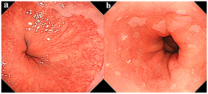
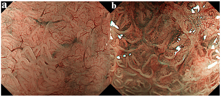
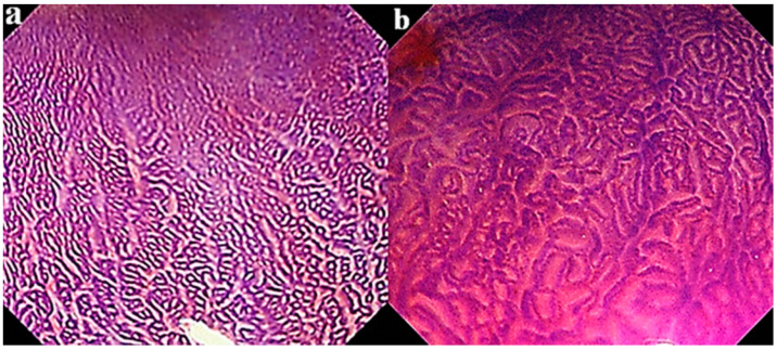
Given that endoscopic findings can be used to predict the potential of neoplastic progression in Barrett's esophagus (BE) cases, the detection rate of dysplastic Barrett's lesions may become higher even in laborious endoscopic surveillance because a special attention is consequently paid. However, endoscopic findings for effective detection of the risk of neoplastic progression to esophageal adenocarcinoma (EAC) have not been confirmed, though some typical appearances are suggestive. In the present review, endoscopic findings that can be used predict malignant potential to EAC in BE cases are discussed. Conventional results obtained with white light endoscopy, such as length of BE, presence of esophagitis, ulceration, hiatal hernia, and nodularity, are used as indicators of a higher risk of neoplastic progression. However, there are controversies in some of those findings. Absence of palisade vessels may be also a new candidate predictor, as that reveals degree of intense inflammation and of cyclooxygenase-2 protein expression with accelerated cellular proliferation. Furthermore, an open type of mucosal pattern and enriched stromal blood vessels, which can be observed by image-enhanced endoscopy, including narrow band imaging, have been confirmed as factors useful for prediction of neoplastic progression of BE because they indicate more frequent cyclooxygenase-2 protein expression along with accelerated cellular proliferation. Should the malignant potential of BE be shown predictable by these endoscopic findings, that would simplify methods used for an effective surveillance, because patients requiring careful monitoring would be more easily identified. Development in the near future of a comprehensive scoring system for BE based on clinical factors, biomarkers and endoscopic predictors is required.
鉴于内镜检查结果可用于预测巴雷特食管(BE)病例的肿瘤进展潜力,即使在内镜检查操作繁琐的情况下,发育异常的巴雷特病变的检出率也可能会更高,因为这样会因此给予特别关注。然而,尽管一些典型表现具有提示性,但尚未证实用于有效检测食管腺癌(EAC)肿瘤进展风险的内镜检查结果。在本综述中,讨论了可用于预测BE病例发生EAC恶性潜能的内镜检查结果。白光内镜检查获得的传统结果,如BE的长度、食管炎、溃疡、食管裂孔疝和结节的存在,被用作肿瘤进展风险较高的指标。然而,其中一些结果存在争议。栅栏状血管缺失也可能是一种新的候选预测指标,因为它揭示了炎症程度以及环氧化酶-2蛋白表达与细胞增殖加速的情况。此外,通过包括窄带成像在内的图像增强内镜观察到的开放型黏膜模式和丰富的基质血管,已被确认为有助于预测BE肿瘤进展的因素,因为它们表明环氧化酶-2蛋白表达更频繁且细胞增殖加速。如果这些内镜检查结果能够显示出可预测BE的恶性潜能,那么将简化有效的监测方法,因为需要仔细监测的患者将更容易被识别。未来需要基于临床因素、生物标志物和内镜预测指标开发一个全面的BE评分系统。