• 文献检索
  • 文档翻译
  • 深度研究
  • 学术资讯
  • Suppr Zotero 插件Zotero 插件
  • 邀请有礼
  • 套餐&价格
  • 历史记录
应用&插件
Suppr Zotero 插件Zotero 插件浏览器插件Mac 客户端Windows 客户端微信小程序
定价
高级版会员购买积分包购买API积分包
服务
文献检索文档翻译深度研究API 文档MCP 服务
关于我们
关于 Suppr公司介绍联系我们用户协议隐私条款
关注我们

Suppr 超能文献

核心技术专利:CN118964589B侵权必究
粤ICP备2023148730 号-1Suppr @ 2026

文献检索

告别复杂PubMed语法,用中文像聊天一样搜索,搜遍4000万医学文献。AI智能推荐,让科研检索更轻松。

立即免费搜索

文件翻译

保留排版,准确专业,支持PDF/Word/PPT等文件格式,支持 12+语言互译。

免费翻译文档

深度研究

AI帮你快速写综述,25分钟生成高质量综述,智能提取关键信息,辅助科研写作。

立即免费体验

33-34 孕周早产儿视网膜未成熟时的视网膜血管发育可预测早产儿视网膜病变。

Retinal vascular development in an immature retina at 33-34 weeks postmenstrual age predicts retinopathy of prematurity.

机构信息

Department of Ophthalmology, Keimyung University School of Medicine, 1095 Dalgebeol-daro, Dalseo-gu, Daegu, 42601, Republic of Korea.

出版信息

Sci Rep. 2020 Oct 22;10(1):18111. doi: 10.1038/s41598-020-75151-0.

DOI:10.1038/s41598-020-75151-0
PMID:33093504
原文链接:https://pmc.ncbi.nlm.nih.gov/articles/PMC7582165/
Abstract

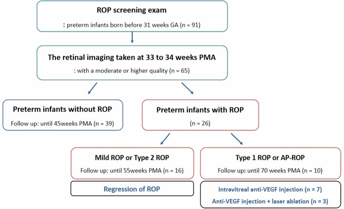
In preterm birth, the immature retina can develop a potentially blinding disorder of the eye known as retinopathy of prematurity (ROP). The vaso-proliferative phase of ROP begins at an approximate postmenstrual age (PMA) of 32 weeks. There is little or no evidence of an association between ROP development and retinal status in the early vaso-proliferative phase. We aimed to evaluate the retinal vascular findings of infants at 33-34 weeks PMA to determine their risk of ROP. We reviewed 130 serial wide-field retinal images from 65 preterm infants born before the gestational age of 31 weeks. ROP occurred more frequently in infants having a leading vascular edge within posterior Zone II. This was in contrast to normal infants, who are characterized by complete retinal vascularization up to Zone II at 34 weeks PMA. The probability of ROP development in preterm infants with retinal edge hemorrhage was 24.58 times higher than in preterm infants without retinal edge hemorrhage. Eyes with ROP that required treatment showed significantly delayed retinal vascularization accompanied by pre-plus disease. In conclusion, retinal status in the early vaso-proliferation phase might determine the risk of ROP.

摘要

在早产儿中,不成熟的视网膜可能会发展出一种称为早产儿视网膜病变(ROP)的潜在致盲眼病。ROP 的血管增生期大约始于孕龄 32 周。ROP 的发展与早期血管增生期视网膜状态之间几乎没有或没有关联的证据。我们旨在评估 33-34 周 PMA 时婴儿的视网膜血管发现,以确定他们患 ROP 的风险。我们回顾了 65 名胎龄小于 31 周的早产儿的 130 张连续广角视网膜图像。ROP 更常发生在具有后二区领先血管边缘的婴儿中。与正常婴儿形成对比,正常婴儿在 34 周 PMA 时视网膜血管化完整延伸到二区。视网膜边缘出血的早产儿发生 ROP 的可能性是没有视网膜边缘出血的早产儿的 24.58 倍。需要治疗的 ROP 眼显示视网膜血管化明显延迟,伴有前加病。总之,早期血管增生期的视网膜状态可能决定 ROP 的风险。

https://cdn.ncbi.nlm.nih.gov/pmc/blobs/b495/7582165/97a7cc90d248/41598_2020_75151_Fig4_HTML.jpg
https://cdn.ncbi.nlm.nih.gov/pmc/blobs/b495/7582165/4cdc3b75e8c7/41598_2020_75151_Fig1_HTML.jpg
https://cdn.ncbi.nlm.nih.gov/pmc/blobs/b495/7582165/4d8fbcb22f40/41598_2020_75151_Fig2_HTML.jpg
https://cdn.ncbi.nlm.nih.gov/pmc/blobs/b495/7582165/83324481b9d9/41598_2020_75151_Fig3_HTML.jpg
https://cdn.ncbi.nlm.nih.gov/pmc/blobs/b495/7582165/97a7cc90d248/41598_2020_75151_Fig4_HTML.jpg

相似文献

1
Retinal vascular development in an immature retina at 33-34 weeks postmenstrual age predicts retinopathy of prematurity.33-34 孕周早产儿视网膜未成熟时的视网膜血管发育可预测早产儿视网膜病变。
Sci Rep. 2020 Oct 22;10(1):18111. doi: 10.1038/s41598-020-75151-0.
2
Maturation of rod function in preterm infants with and without retinopathy of prematurity.患有和未患早产儿视网膜病变的早产儿视杆功能成熟情况。
J Pediatr. 2008 Nov;153(5):605-11. doi: 10.1016/j.jpeds.2008.05.018. Epub 2008 Jul 14.
3
RATE OF AND TIME TO COMPLETE RETINAL VASCULARIZATION IN PREMATURE INFANTS AND ASSOCIATED FACTORS.早产儿视网膜血管化完成的速率及时间与相关因素
Retina. 2023 Jan 1;43(1):102-110. doi: 10.1097/IAE.0000000000003627.
4
Management of avascular peripheral retina in older prematurely born infants.早产儿老龄患者外周无血管视网膜的处理
Retina. 2011 Jul-Aug;31(7):1248-54. doi: 10.1097/IAE.0b013e31820d3f70.
5
Integrated Visualization Highlighting Retinal Changes in Retinopathy of Prematurity From 3-Dimensional Optical Coherence Tomography Data.基于三维光学相干断层扫描数据的综合可视化突出早产儿视网膜病变的视网膜变化。
JAMA Ophthalmol. 2022 Jul 1;140(7):725-729. doi: 10.1001/jamaophthalmol.2022.1344.
6
Intraocular Hemorrhages and Retinopathy of Prematurity in the Telemedicine Approaches to Evaluating Acute-Phase Retinopathy of Prematurity (e-ROP) Study.远程医疗评估早产儿急性视网膜病变(e-ROP)研究中的眼内出血与早产儿视网膜病变
Ophthalmology. 2017 Mar;124(3):374-381. doi: 10.1016/j.ophtha.2016.10.040. Epub 2016 Dec 13.
7
Zone of retinal vascularization and refractive error in premature eyes with and without spontaneously regressed retinopathy of prematurity.患有和未患有自然消退型早产儿视网膜病变的早产眼的视网膜血管化区域和屈光不正
J AAPOS. 2019 Aug;23(4):211.e1-211.e6. doi: 10.1016/j.jaapos.2019.03.006. Epub 2019 Jun 20.
8
Retinopathy of prematurity in infants born before 25 weeks gestation in a Korean single neonatal intensive care unit: incidence, natural history and risk factors.韩国单家新生儿重症监护病房 25 周以下早产儿早产儿视网膜病变:发生率、自然病程和危险因素。
J Korean Med Sci. 2012 Dec;27(12):1556-62. doi: 10.3346/jkms.2012.27.12.1556. Epub 2012 Dec 7.
9
Natural course of retinal development in preterm infants without threshold retinopathy.无阈值视网膜病变的早产儿视网膜发育的自然病程
Ger J Ophthalmol. 1995 May;4(3):131-6.
10
Delayed resolution of retinopathy of prematurity.早产儿视网膜病变的延迟消退。
J AAPOS. 2019 Apr;23(2):90.e1-90.e6. doi: 10.1016/j.jaapos.2018.10.012. Epub 2019 Feb 14.

引用本文的文献

1
The Long-Standing Problem of Proliferative Retinopathies: Current Understanding and Critical Cues.增殖性视网膜病变的长期问题:当前认识与关键线索
Cells. 2025 Jul 18;14(14):1107. doi: 10.3390/cells14141107.
2
Evaluation of the Course and Outcome of Aggressive Retinopathy of Prematurity.早产儿侵袭性视网膜病变的病程及预后评估
Cureus. 2025 May 21;17(5):e84589. doi: 10.7759/cureus.84589. eCollection 2025 May.
3
A retrospective study of ophthalmologic presentation, management, and outcomes in pediatric patients admitted with abusive head trauma.

本文引用的文献

1
Imaging in Retinopathy of Prematurity.早产儿视网膜病变的影像学检查。
Asia Pac J Ophthalmol (Phila). 2019 Mar-Apr;8(2):178-186. doi: 10.22608/APO.201963.
2
Comparison of fluorescein angiographic findings in type 1 and type 2 retinopathy of prematurity with intravitreal bevacizumab monotherapy and spontaneous regression.1型和2型早产儿视网膜病变接受玻璃体内注射贝伐单抗单药治疗及自然消退后的荧光素血管造影结果比较
Int Ophthalmol. 2019 Oct;39(10):2267-2274. doi: 10.1007/s10792-018-01064-7. Epub 2019 Jan 2.
3
Spectral-Domain OCT Findings of Retinal Vascular-Avascular Junction in Infants with Retinopathy of Prematurity.
对因虐待性头部外伤入院的儿科患者的眼科表现、治疗及预后的回顾性研究。
Front Med (Lausanne). 2024 Aug 15;11:1416626. doi: 10.3389/fmed.2024.1416626. eCollection 2024.
4
Changes of the peripapillary vascular parameters in premature infants without retinopathy of prematurity using U-net segmentation.使用U-net分割技术对无早产儿视网膜病变的早产儿视乳头周围血管参数的变化
Int J Ophthalmol. 2024 Aug 18;17(8):1453-1461. doi: 10.18240/ijo.2024.08.10. eCollection 2024.
5
The Role of HIF-1α in Retinopathy of Prematurity: A Review of Current Literature.缺氧诱导因子-1α在早产儿视网膜病变中的作用:当前文献综述
J Clin Med. 2024 Jul 10;13(14):4034. doi: 10.3390/jcm13144034.
6
Mechanical Ventilation, Retinal Avascularity and Rate of Vascularisation: A Triad of Predictors for Retinopathy of Prematurity Treatment.机械通气、视网膜无血管化与血管化速率:早产儿视网膜病变治疗的三联预测指标
J Pers Med. 2024 Mar 31;14(4):379. doi: 10.3390/jpm14040379.
7
Early Single-Examination Optical Coherence Tomography Biomarkers for Treatment-Requiring Retinopathy of Prematurity.早期单检查光学相干断层扫描生物标志物预测早产儿视网膜病变需治疗的情况
Invest Ophthalmol Vis Sci. 2024 Apr 1;65(4):21. doi: 10.1167/iovs.65.4.21.
8
The incidence of retinopathy of prematurity in neonates in Germany in 2019; a nationwide epidemiological cohort study.2019年德国新生儿早产儿视网膜病变的发病率;一项全国性的流行病学队列研究。
Eur J Pediatr. 2024 Feb;183(2):827-834. doi: 10.1007/s00431-023-05229-0. Epub 2023 Nov 30.
9
Primary and additional treatment preference in aggressive retinopathy of prematurity and type 1 retinopathy of prematurity.早产儿视网膜病变和 1 型早产儿视网膜病变的主要和辅助治疗偏好。
BMJ Open Ophthalmol. 2023 Jan;8(1). doi: 10.1136/bmjophth-2022-001166.
10
Cord blood transforming growth factor-β-induced as predictive biomarker of retinopathy of prematurity in preterm infants.脐带血转化生长因子-β诱导作为早产儿视网膜病变的预测生物标志物。
Graefes Arch Clin Exp Ophthalmol. 2023 Sep;261(9):2477-2488. doi: 10.1007/s00417-023-06056-7. Epub 2023 Apr 6.
早产儿视网膜病变婴儿视网膜血管-无血管交界处的频域光学相干断层扫描结果
Ophthalmol Retina. 2018 Sep;2(9):963-971. doi: 10.1016/j.oret.2018.02.001. Epub 2018 Mar 21.
4
Incidence and Early Course of Retinopathy of Prematurity: Secondary Analysis of the Postnatal Growth and Retinopathy of Prematurity (G-ROP) Study.早产儿视网膜病变的发生率和早期病程:产后生长与早产儿视网膜病变(G-ROP)研究的二次分析。
JAMA Ophthalmol. 2018 Dec 1;136(12):1383-1389. doi: 10.1001/jamaophthalmol.2018.4290.
5
Retinal Vasculature in Development and Diseases.视网膜血管在发育和疾病中的作用
Annu Rev Vis Sci. 2018 Sep 15;4:101-122. doi: 10.1146/annurev-vision-091517-034018.
6
Plus Disease in Retinopathy of Prematurity: More Than Meets the ICROP?早产儿视网膜病变中的 Plus 病:比 ICROP 所见更复杂?
Asia Pac J Ophthalmol (Phila). 2018 May-Jun;7(3):152-155. doi: 10.22608/APO.201863. Epub 2018 May 24.
7
An Update on Retinopathy of Prematurity (ROP).早产儿视网膜病变(ROP)的最新进展。
Indian J Pediatr. 2017 Dec;84(12):930-936. doi: 10.1007/s12098-017-2404-3. Epub 2017 Jul 4.
8
RETINAL VASCULAR DEVELOPMENT WITH 0.312 MG INTRAVITREAL BEVACIZUMAB TO TREAT SEVERE POSTERIOR RETINOPATHY OF PREMATURITY: A Longitudinal Fluorescein Angiographic Study.玻璃体内注射0.312毫克贝伐单抗治疗重度早产儿视网膜病变的视网膜血管发育:一项纵向荧光素血管造影研究
Retina. 2017 Jan;37(1):97-111. doi: 10.1097/IAE.0000000000001126.
9
Embryologic and Fetal Development of the Human Eyelid.人类眼睑的胚胎学和胎儿发育
Ophthalmic Plast Reconstr Surg. 2016 Nov/Dec;32(6):407-414. doi: 10.1097/IOP.0000000000000702.
10
Do we screen very premature babies too early for retinopathy of prematurity?我们对极早产儿筛查早产儿视网膜病变是否为时过早?
Eye (Lond). 2016 Apr;30(4):636-7. doi: 10.1038/eye.2015.279. Epub 2016 Jan 22.