Wilt Haley M, Yu Ping, Tan Kemin, Wang Yun-Xing, Stagno Jason R
Structural Biophysics Laboratory, Center for Cancer Research, National Cancer Institute, Frederick, MD 21702, USA.
Washington College, Chestertown, Maryland 21620, USA.
J Struct Biol X. 2020 Aug 6;4:100035. doi: 10.1016/j.yjsbx.2020.100035. eCollection 2020.
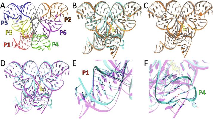
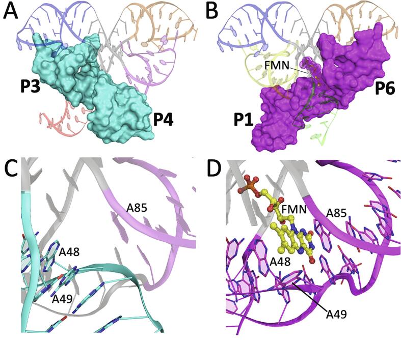
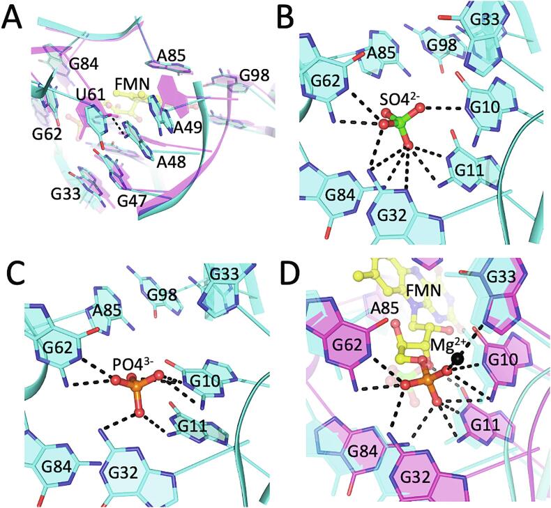
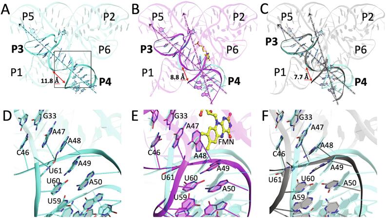
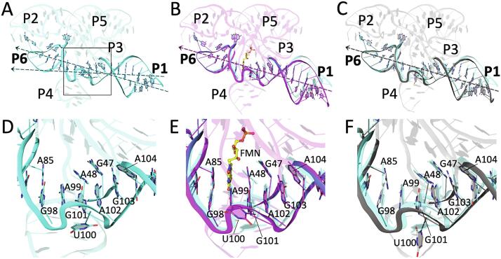
Knowledge of both apo and holo states of riboswitches aid in elucidating the various mechanisms of ligand-induced conformational "switching" that underpin their gene-regulating capabilities. Previous structural studies on the flavin mononucleotide (FMN)-binding aptamer of the FMN riboswitch, however, have revealed minimal conformational changes associated with ligand binding that do not adequately explain the basis for the switching behavior. We have determined a 2.7-Å resolution crystal structure of the ligand-free FMN riboswitch aptamer that is distinct from previously reported structures, particularly in the conformation and orientation of the P1 and P4 helices. The nearly symmetrical tertiary structure provides a mechanism by which one of two pairs of adjacent helices (P3/P4 or P1/P6) undergo collinear stacking in a mutually exclusive manner, in the absence or presence of ligand, respectively. Comparison of these structures suggests the stem-loop that includes P4 and L4 is important for maintaining a global conformational state that, in the absence of ligand, disfavors formation of the P1 regulatory helix. Together, these results provide further insight to the structural basis for conformational switching of the FMN riboswitch.
对核糖开关的脱辅基和全酶状态的了解有助于阐明配体诱导的构象“转换”的各种机制,这些机制是其基因调控能力的基础。然而,先前对黄素单核苷酸(FMN)核糖开关的FMN结合适体的结构研究表明,与配体结合相关的构象变化极小,不足以解释开关行为的基础。我们确定了无配体的FMN核糖开关适体的2.7埃分辨率晶体结构,该结构与先前报道的结构不同,特别是在P1和P4螺旋的构象和取向上。近乎对称的三级结构提供了一种机制,通过该机制,两对相邻螺旋(P3/P4或P1/P6)中的一对分别在无配体或有配体的情况下以互斥方式进行共线堆积。这些结构的比较表明,包括P4和L4的茎环对于维持整体构象状态很重要,在无配体的情况下,这种构象状态不利于P1调节螺旋的形成。总之,这些结果为FMN核糖开关构象转换的结构基础提供了进一步的见解。