Bryja Artur, Sujka-Kordowska Patrycja, Konwerska Aneta, Ciesiółka Sylwia, Wieczorkiewicz Maria, Bukowska Dorota, Antosik Paweł, Bryl Rut, Skowroński Mariusz T, Jaśkowski Jędrzej M, Mozdziak Paul, Angelova Volponi Ana, Shibli Jamil A, Kempisty Bartosz, Dyszkiewicz-Konwińska Marta
Department of Anatomy, Poznan University of Medical Science, 60-781 Poznań, Poland.
Department of Histology and Embryology, Poznan University of Medical Science, 60-781 Poznań, Poland.
Animals (Basel). 2020 Oct 22;10(11):1938. doi: 10.3390/ani10111938.
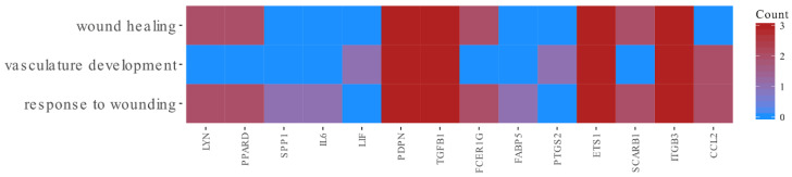
The mechanisms of wound healing and vascularization are crucial steps of the complex morphological process of tissue reconstruction. In addition to epithelial cells, fibroblasts play an important role in this process. They are characterized by dynamic proliferation and they form the stroma for epithelial cells. In this study, we have used primary cultures of oral fibroblasts, obtained from porcine buccal mucosa. Cells were maintained long-term in in vitro conditions, in order to investigate the expression profile of the molecular markers involved in wound healing and vascularization. Based on the Affymetrix assays, we have observed three ontological groups of markers as , and , represented by different genes characterized by their expression profile during long-term primary in vitro culture (IVC) of porcine oral fibroblasts. Following the analysis of gene expression in three previously identified groups of genes, we have identified that transforming growth factor beta 1 (), , , and are involved in all three processes, suggesting that these genes could be recognized as markers of repair specific for oral fibroblasts within the porcine mucosal tissue.
伤口愈合和血管生成机制是组织重建这一复杂形态学过程的关键步骤。除上皮细胞外,成纤维细胞在此过程中也发挥着重要作用。它们的特点是动态增殖,并为上皮细胞形成基质。在本研究中,我们使用了从猪颊黏膜获取的原代口腔成纤维细胞培养物。细胞在体外条件下长期培养,以研究参与伤口愈合和血管生成的分子标志物的表达谱。基于Affymetrix分析,我们观察到三类本体标志物,即A、B和C,它们由不同基因代表,这些基因的特征在于其在猪口腔成纤维细胞长期原代体外培养(IVC)过程中的表达谱。在分析先前确定的三组基因中的基因表达后,我们发现转化生长因子β1(TGF-β1)、A、B和C参与了所有三个过程,这表明这些基因可被视为猪黏膜组织中口腔成纤维细胞特异性修复的标志物。