Qiu Yao, Sun Lizhi, Hu Xiaolin, Zhao Xin, Shi Hongyan, Liu Zhao, Yin Xiao
Department of Surgery, Shandong University Affiliated Jinan Central Hospital, Jinan, Shandong People's Republic of China.
Department of Clinical Laboratory, Shandong University Affiliated Jinan Central Hospital, Jinan, Shandong People's Republic of China.
Diabetol Metab Syndr. 2020 Oct 22;12:91. doi: 10.1186/s13098-020-00599-z. eCollection 2020.
People with obesity have a compromised browning capacity of adipose tissue when faced with sympathetic stimuli. This study aimed to determine whether norepinephrine treatment can enhance the induction of precursor cells from human white adipose tissue to differentiate into adipocytes that express key markers of beige adipocytes, and if there is a difference in this capacity between normal weight and overweight individuals.
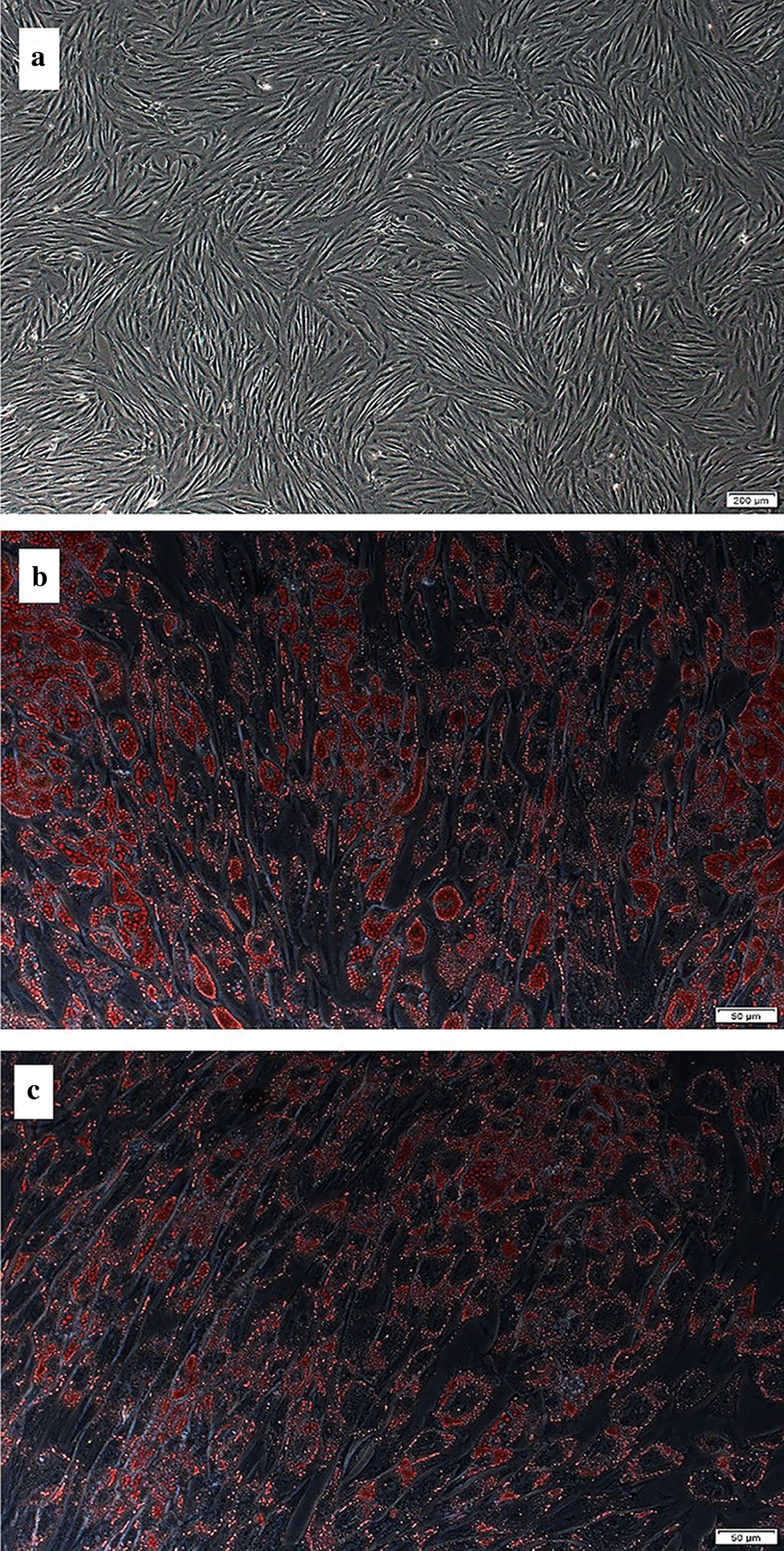
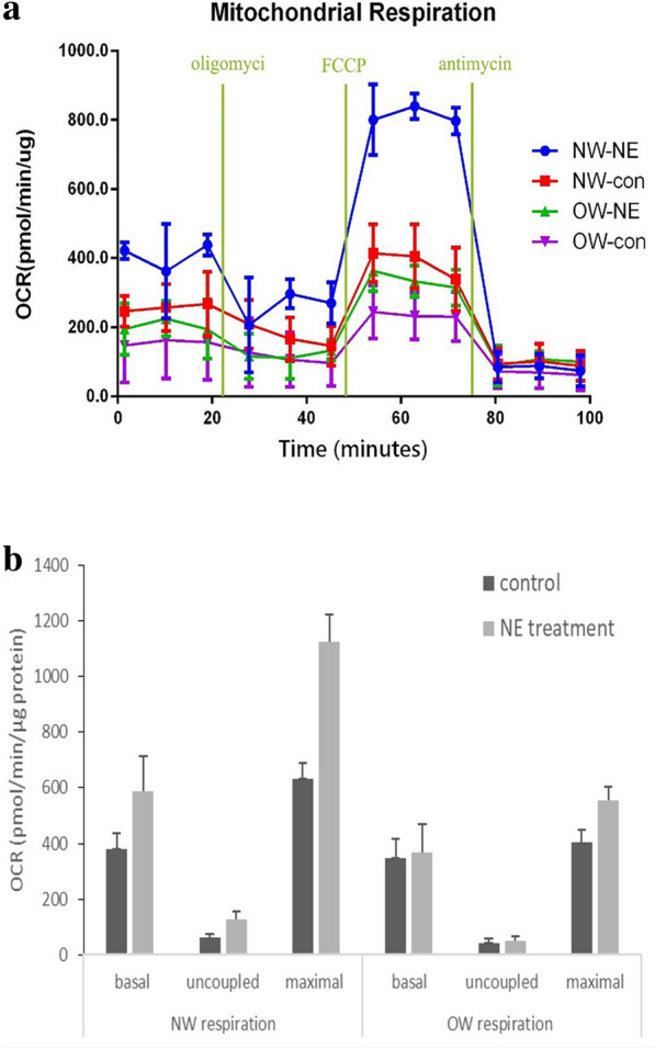
Stromal vascular cells derived from subcutaneous white adipose tissue of normal weight and overweight groups were induced to differentiation, with or without norepinephrine, into adipocytes. Oxygen consumption rate, lipolysis, the expression of uncoupling protein 1 and other thermogenic genes were compared between different adiposity and treatment groups.
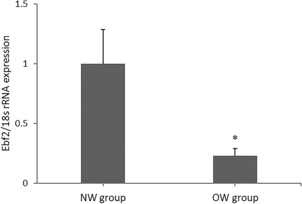
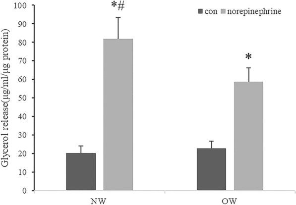
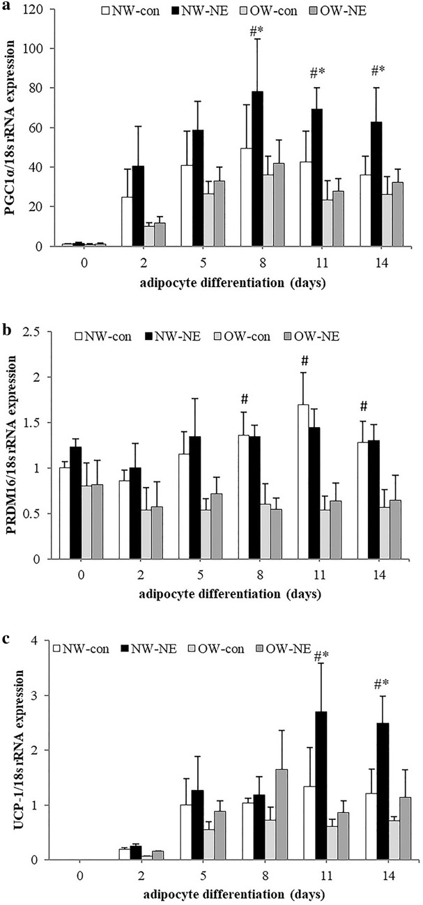
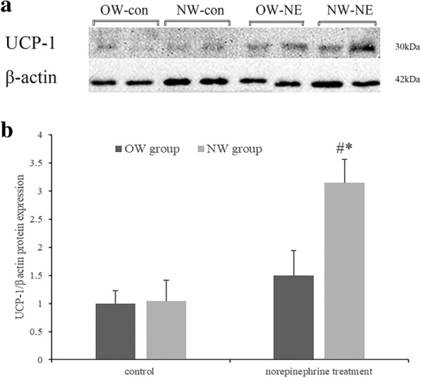
Peroxisome proliferator activated receptor γ- coactivator-1 alpha (PGC-1 α) and uncoupling protein 1 gene expression increased significantly in the normal weight group, but not in the overweight group, with norepinephrine treatment. The increments of lipolysis and oxygen consumption rate were also higher in adipocytes from the normal weight group with norepinephrine treatment, as compared with those of the overweight group. PR domain containing protein 16 (PRDM 16) gene expression was higher in the normal weight group compared with that in the overweight group, while there were no significant changes found with norepinephrine treatment in either the normal weight or overweight group.
Adipogenic precursor cells derived from overweight individuals were less prone to differentiate into beige-like adipocytes when facing sympathetic stimuli than normal weight ones, resulting in the compromised sympathetic-induced browning capacity in subcutaneous white adipose tissue in overweight individuals, which occurred before the onset of overt obesity.
肥胖人群在面对交感神经刺激时,脂肪组织的褐色化能力受损。本研究旨在确定去甲肾上腺素治疗是否能增强人白色脂肪组织中前体细胞向表达米色脂肪细胞关键标志物的脂肪细胞分化的诱导作用,以及正常体重和超重个体在这种能力上是否存在差异。
将正常体重组和超重组皮下白色脂肪组织来源的基质血管细胞在有或没有去甲肾上腺素的情况下诱导分化为脂肪细胞。比较不同肥胖程度和治疗组之间的氧消耗率、脂肪分解、解偶联蛋白1及其他产热基因的表达。
在去甲肾上腺素治疗下,正常体重组中过氧化物酶体增殖物激活受体γ-辅激活因子-1α(PGC-1α)和解偶联蛋白1基因表达显著增加,而超重组则未增加。与超重组相比,去甲肾上腺素治疗的正常体重组脂肪细胞的脂肪分解增加量和氧消耗率也更高。正常体重组中含PR结构域蛋白16(PRDM16)基因表达高于超重组,而去甲肾上腺素治疗在正常体重组或超重组中均未发现显著变化。
超重个体来源的脂肪生成前体细胞在面对交感神经刺激时比正常体重个体更不易分化为米色样脂肪细胞,导致超重个体皮下白色脂肪组织中交感神经诱导的褐色化能力受损,这在明显肥胖发生之前就已出现。