9100 Babcock Boulevard, Professional Building T, Pittsburgh, PA, 15237, United States.
Department of Neurology, Baylor College of Medicine, Houston, TX, 77030, United States; Neurology Care Line, VA Houston Medical Center, Houston, TX, 77030, United States.
Seizure. 2020 Dec;83:234-241. doi: 10.1016/j.seizure.2020.10.014. Epub 2020 Oct 19.
We performed a systematic review of the literature to synthesize the data on EEG findings in COVID-19. Frontal EEG patterns are reported to be a characteristic finding in COVID-19 encephalopathy. Although several reports of EEG abnormalities are available, there is lack of clarity about typical findings.
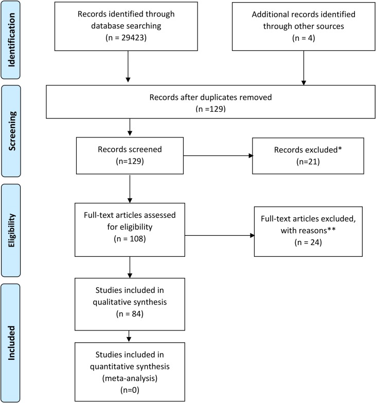
Research databases were queried with the terms "COVID" OR "coronavirus" OR "SARS" AND "EEG". Available data was analyzed from 617 patients with EEG findings reported in 84 studies.
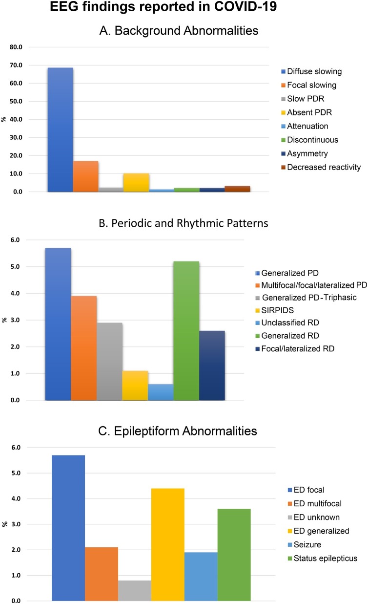
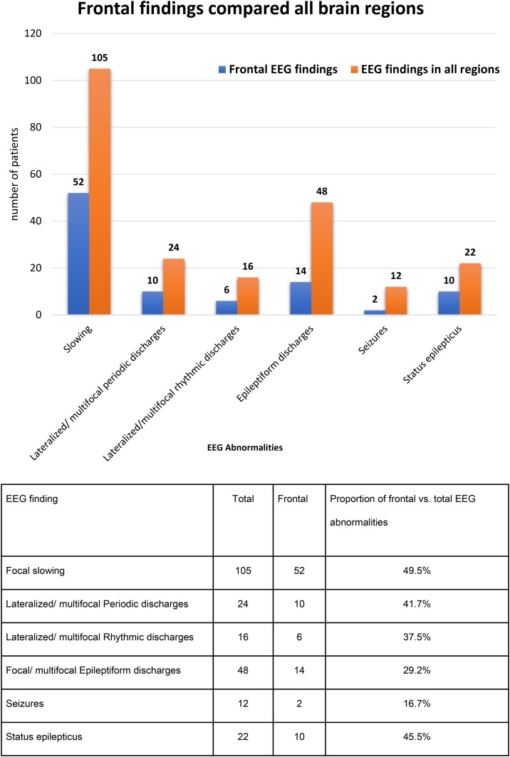
The median age was 61.3 years (IQR 45-69, 33.3 % female). Common EEG indications were altered mental status (61.7 %), seizure-like events (31.2 %), and cardiac arrest (3.5 %). Abnormal EEG findings (n = 543, 88.0 %) were sub-classified into three groups: (1) Background abnormalities: diffuse slowing (n = 423, 68.6 %), focal slowing (n = 105, 17.0 %), and absent posterior dominant rhythm (n = 63, 10.2 %). (2) Periodic and rhythmic EEG patterns: generalized periodic discharges (n = 35, 5.7 %), lateralized/multifocal periodic discharges (n = 24, 3.9 %), generalized rhythmic activity (n = 32, 5.2 %). (3) Epileptiform changes: focal (n = 35, 5.7 %), generalized (n = 27, 4.4 %), seizures/status epilepticus (n = 34, 5.5 %). Frontal EEG patterns comprised of approximately a third of all findings. In studies that utilized continuous EEG, 96.8 % (n = 243) of the 251 patients were reported to have abnormalities compared to 85.0 % (n = 311) patients who did not undergo continuous EEG monitoring (χ = 22.8, p =< 0.001).
EEG abnormalities are common in COVID-19 related encephalopathy and correlates with disease severity, preexisting neurological conditions including epilepsy and prolonged EEG monitoring. Frontal findings are frequent and have been proposed as a biomarker for COVID-19 encephalopathy.
我们对文献进行了系统综述,以综合 COVID-19 患者脑电图发现的数据。据报道,额叶脑电图模式是 COVID-19 脑病的特征性发现。尽管有几项关于脑电图异常的报告,但对于典型发现尚缺乏明确性。
用“COVID”或“冠状病毒”或“SARS”和“EEG”等术语查询研究数据库。对 84 项研究中报告的 617 例脑电图结果进行数据分析。
中位年龄为 61.3 岁(IQR 45-69,33.3%为女性)。常见的脑电图指征包括意识状态改变(61.7%)、癫痫样事件(31.2%)和心搏骤停(3.5%)。异常脑电图发现(n=543,88.0%)分为三组:(1)背景异常:弥漫性减慢(n=423,68.6%)、局灶性减慢(n=105,17.0%)和后部优势节律缺失(n=63,10.2%)。(2)周期性和节律性脑电图模式:全面性周期性放电(n=35,5.7%)、偏侧性/多灶性周期性放电(n=24,3.9%)、全面性节律性活动(n=32,5.2%)。(3)癫痫样改变:局灶性(n=35,5.7%)、全面性(n=27,4.4%)、癫痫发作/癫痫持续状态(n=34,5.5%)。额叶脑电图模式约占所有发现的三分之一。在使用连续脑电图的研究中,与未行连续脑电图监测的患者(n=311,85.0%)相比,96.8%(n=243)的 251 例患者报告有异常(χ²=22.8,p<0.001)。
脑电图异常在 COVID-19 相关脑病中很常见,与疾病严重程度、包括癫痫在内的先前存在的神经疾病以及延长的脑电图监测相关。额叶发现很常见,已被提议作为 COVID-19 脑病的生物标志物。