Radiology Unit, Department of Pathology and Diagnostics, Azienda Ospedaliera Universitaria Integrata - Verona, P.le Stefani 1, 37126, Verona, Italy.
Medical Physics Unit, Department of Pathology and Diagnostics, Azienda Ospedaliera Universitaria Integrata - Verona, P.le Stefani 1, 37126, Verona, Italy.
Eur Radiol. 2021 May;31(5):2645-2656. doi: 10.1007/s00330-020-07396-2. Epub 2020 Oct 30.
This study evaluated the feasibility of DWI for lesion targeting in MRI-guided breast biopsies. Furthermore, it assessed device positioning on DWI during biopsy procedures.
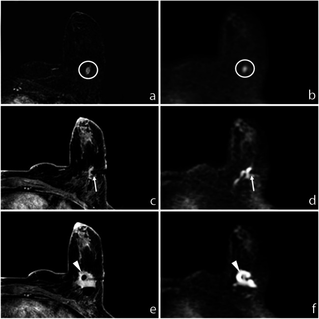
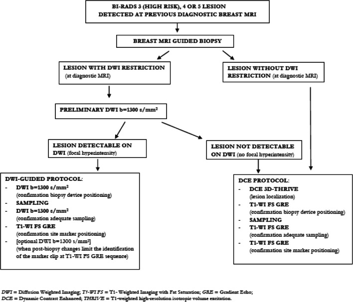
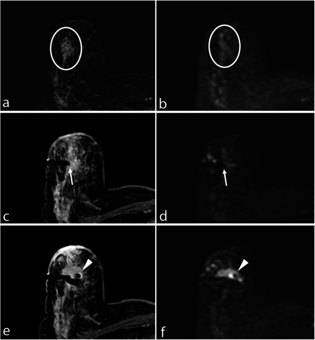
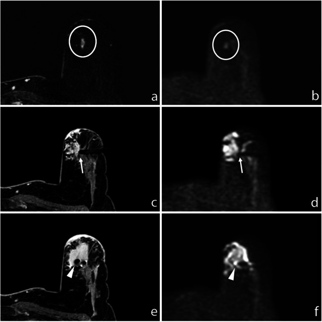
A total of 87 biopsy procedures (5/87 bilateral) consecutively performed between March 2019 and June 2020 were retrospectively reviewed: in these procedures, a preliminary DWI sequence (b = 1300 s/mm) was acquired to assess lesion detectability. We included 64/87 procedures on lesions detectable at DWI; DWI sequences were added to the standard protocol to localize lesion and biopsy device and to assess the site marker correct positioning.
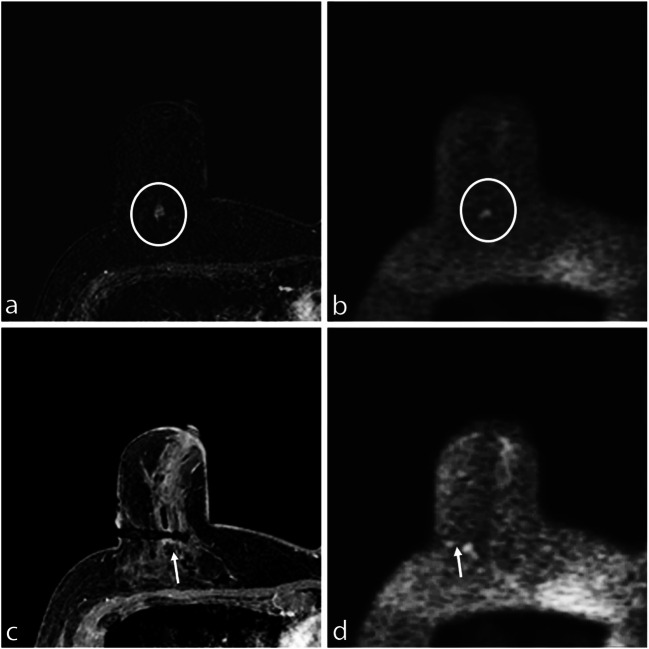
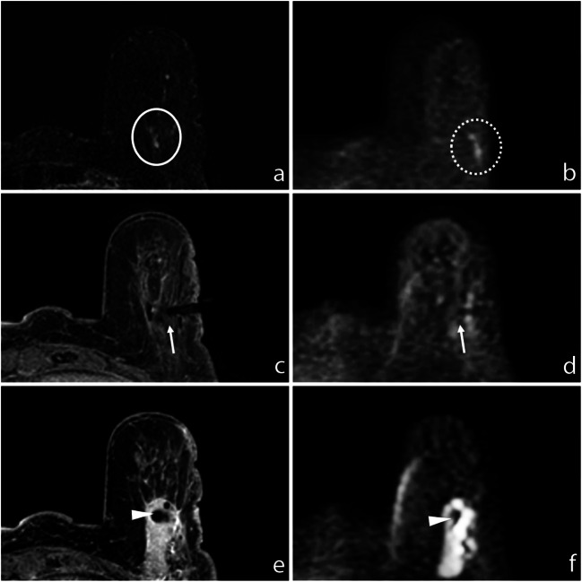
Mass lesions ranged from 5 to 48 mm, with a mean size of 10.7 mm and a median size of 8 mm. Non-mass lesions ranged from 7 to 90 mm, with a mean size of 33.9 mm and a median size of 31 mm. Positioning of the coaxial system was confirmed on both T1-weighted and DWI sequences. At DWI, the biopsy needle was detectable in 62/64 (96.9%) cases; it was not visible in 2/64 (3.1%) cases. The site marker was always identified using T1-weighted imaging; a final DWI sequence was acquired in 44/64 cases (68.8%). In 42/44 cases (95.5%), the marker was recognizable at DWI.
DWI can be used as a cost-effective, highly reliable technique for targeting both mass and non-mass lesions, with a minimum size of 5 mm, detectable at pre-procedural DWI. DWI is also a feasible technique to localize the biopsy device and to confirm the deployment of the site marker.
• MRI-guided breast biopsy is performed in referral centers by an expert dedicated staff, based on prior MR imaging; contrast agent administration is usually needed for lesion targeting. • DWI represents a feasible, highly reliable technique for lesion targeting, avoiding contrast agent administration. • DWI allows a precise localization of both biopsy needle device and site marker.
本研究评估了磁共振引导下乳腺活检中弥散加权成像(DWI)用于病灶定位的可行性,并评估了在活检过程中设备在 DWI 上的定位。
回顾性分析 2019 年 3 月至 2020 年 6 月连续进行的 87 例(5/87 例双侧)活检手术:这些手术中,首先进行初步 DWI 序列(b=1300 s/mm)以评估病灶的可检测性。我们纳入了 64/87 例可在 DWI 上检测到的病灶的手术,DWI 序列被添加到标准方案中,以定位病灶和活检设备,并评估部位标记物的正确定位。
肿块病变的范围从 5 到 48mm,平均大小为 10.7mm,中位数为 8mm。非肿块病变的范围从 7 到 90mm,平均大小为 33.9mm,中位数为 31mm。同轴系统的定位在 T1 加权和 DWI 序列上均得到了确认。在 DWI 上,62/64(96.9%)例活检针可检测到,2/64(3.1%)例不可检测到。部位标记物始终可以通过 T1 加权成像识别,在 44/64 例(68.8%)中最终获得了 DWI 序列。在 42/44 例(95.5%)中,标记物在 DWI 上可识别。
DWI 可以作为一种经济有效的、高度可靠的技术,用于定位大小至少为 5mm 的肿块和非肿块病变,在术前 DWI 上即可检测到。DWI 也是一种用于定位活检设备和确认部位标记物部署的可行技术。
MRI 引导下乳腺活检由专门的专家团队在转诊中心进行,基于术前磁共振成像;病灶定位通常需要使用造影剂。
DWI 是一种可行的、高度可靠的病灶定位技术,可避免造影剂的使用。
DWI 可以精确地定位活检针设备和部位标记物。