Department of Biological Chemistry, The Alexander Silberman Institute of Life Sciences, The Hebrew University of Jerusalem, Jerusalem 9190401, Israel.
Bio-Imaging Unit, The Alexander Silberman Institute of Life Sciences, The Hebrew University of Jerusalem, Jerusalem 9190401, Israel.
J Mol Cell Cardiol. 2021 Jun;155:125-137. doi: 10.1016/j.yjmcc.2020.10.013. Epub 2020 Oct 30.
One unaddressed aspect of healing after myocardial infarction (MI) is how non-myocyte cells that survived the ischemic injury, keep withstanding additional cellular damage by stress forms typically arising during the post-infarction inflammation. Here we aimed to determine if cell survival is conferred by expression of a mitochondrial protein novel to the cardiac proteome, known as steroidogenic acute regulatory protein, (StAR/STARD1). Further studies aimed to unravel the regulation and role of the non-steroidogenic cardiac StAR after MI.
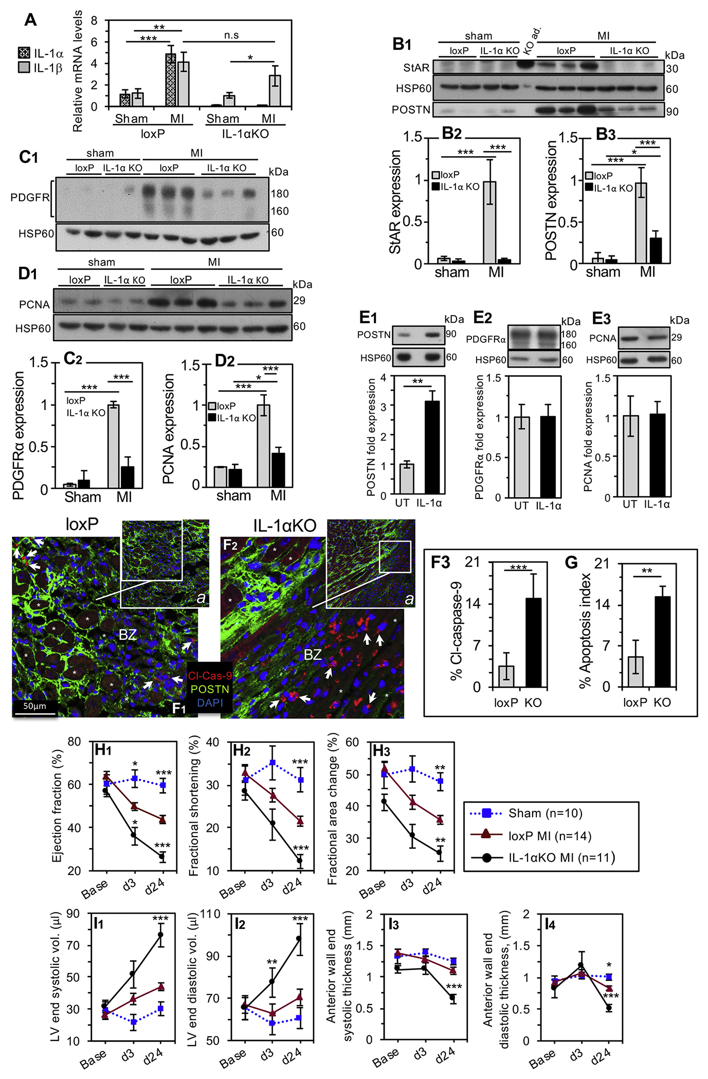
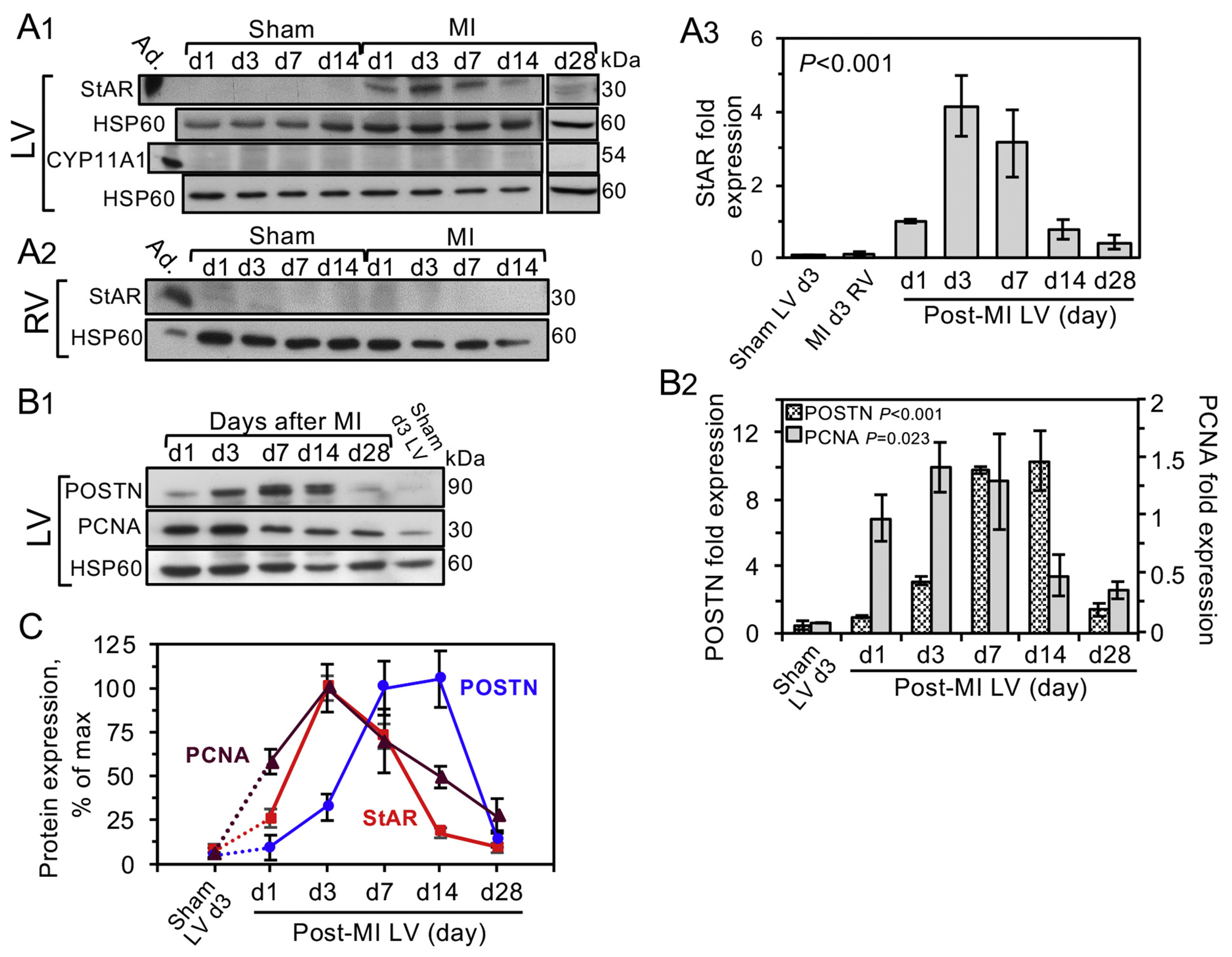
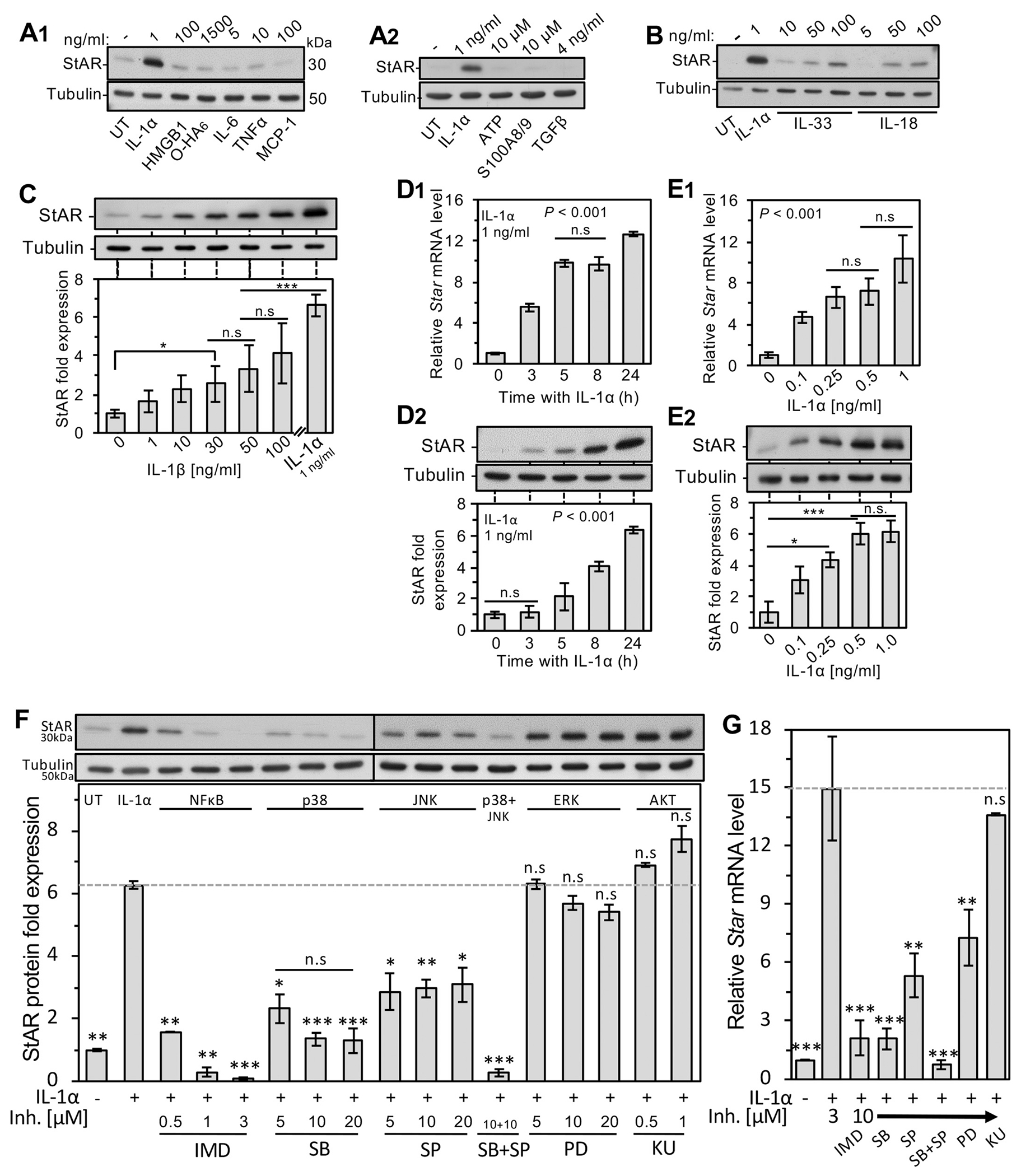
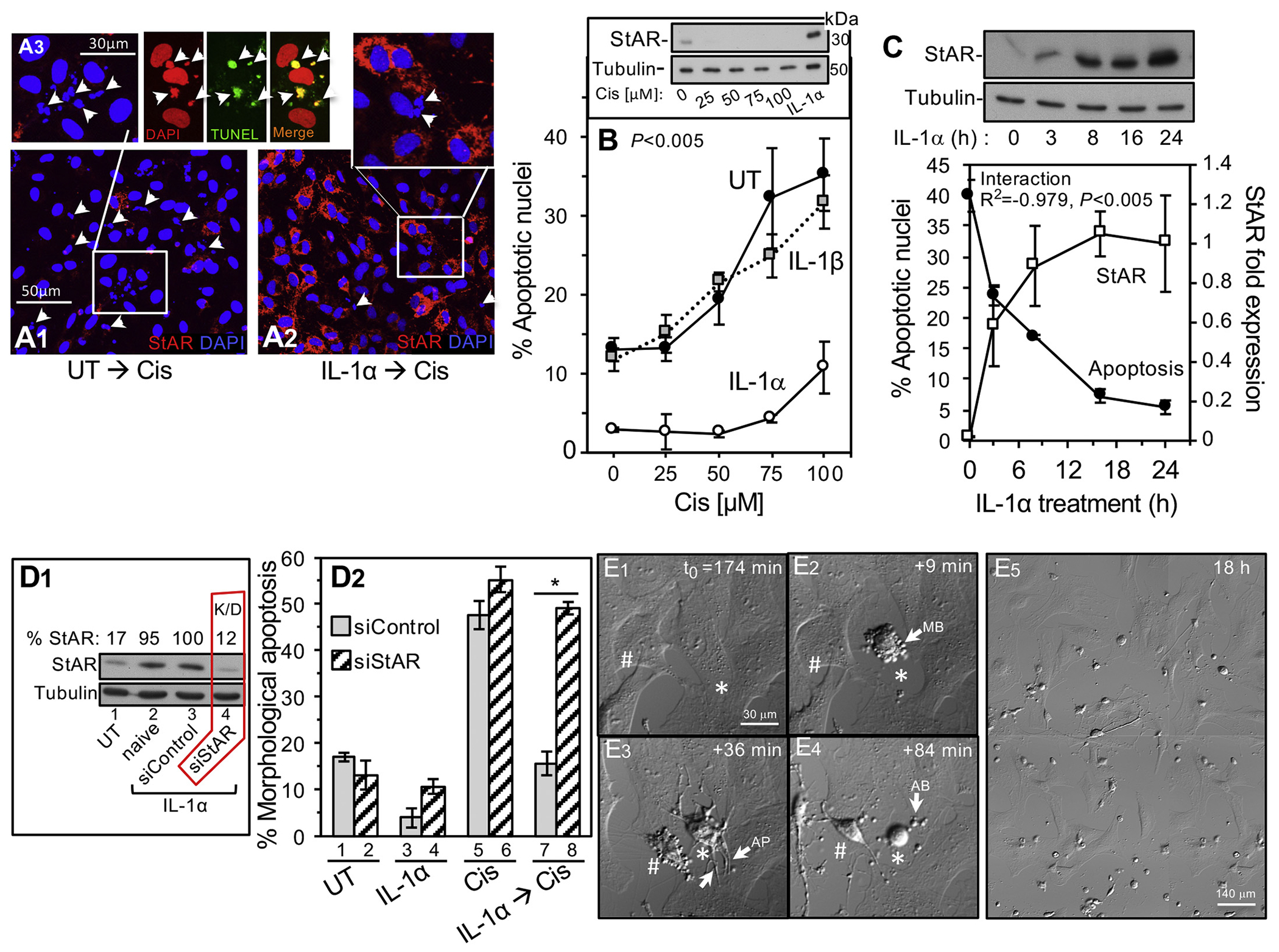
Following permanent ligation of the left anterior descending coronary artery in mouse heart, timeline western blot analyses showed that StAR expression corresponds to the inflammatory response to MI. Following the identification of StAR in mitochondria of cardiac fibroblasts in culture, confocal microscopy immunohistochemistry (IHC) identified StAR expression in left ventricular (LV) activated interstitial fibroblasts, adventitial fibroblasts and endothelial cells. Further work with the primary fibroblasts model revealed that interleukin-1α (IL-1α) signaling via NF-κB and p38 MAPK pathways efficiently upregulates the expression of the Star gene products. At the functional level, IL-1α primed fibroblasts were protected against apoptosis when exposed to cisplatin mimicry of in vivo apoptotic stress; yet, the protective impact of IL-1α was lost upon siRNA mediated StAR downregulation. At the physiological level, StAR expression was nullified during post-MI inflammation in a mouse model with global IL-1α deficiency, concomitantly resulting in a 4-fold elevation of apoptotic fibroblasts. Serial echocardiography and IHC studies of mice examined 24 days after MI revealed aggravation of LV dysfunction, LV dilatation, anterior wall thinning and adverse tissue remodeling when compared with loxP control hearts.
This study calls attention to overlooked aspects of cellular responses evolved under the stress conditions associated with the default inflammatory response to MI. Our observations suggest that LV IL-1α is cardioprotective, and at least one mechanism of this action is mediated by induction of StAR expression in border zone fibroblasts, which renders them apoptosis resistant. This acquired survival feature also has long-term ramifications on the heart recovery by diminishing adverse remodeling and improving the heart function after MI.
心肌梗死 (MI) 后愈合的一个未解决的问题是,在梗死后炎症期间通常出现的应激形式下,幸存下来的非心肌细胞如何继续承受额外的细胞损伤。在这里,我们旨在确定是否通过表达一种新的心脏蛋白质组中的线粒体蛋白,即类固醇急性调节蛋白(StAR/STARD1)来赋予细胞存活。进一步的研究旨在阐明 MI 后非甾体心脏 StAR 的调节和作用。
在小鼠心脏左前降支永久结扎后,时间进程 Western blot 分析表明,StAR 的表达与 MI 的炎症反应相对应。在鉴定出培养的心脏成纤维细胞中的 StAR 存在于线粒体中后,共聚焦显微镜免疫组化(IHC)鉴定出左心室(LV)激活的间质成纤维细胞、外膜成纤维细胞和内皮细胞中的 StAR 表达。使用原代成纤维细胞模型的进一步研究表明,白细胞介素 1α(IL-1α)通过 NF-κB 和 p38 MAPK 途径的信号转导有效地上调了 Star 基因产物的表达。在功能水平上,当暴露于体内凋亡应激的顺铂模拟物时,IL-1α 预先刺激的成纤维细胞被保护免于凋亡;然而,当使用 siRNA 介导的 StAR 下调时,IL-1α 的保护作用丧失。在生理水平上,在缺乏全身性 IL-1α 的小鼠模型中,在梗死后炎症期间,StAR 表达被消除,同时凋亡成纤维细胞的数量增加了 4 倍。对 MI 后 24 天的小鼠进行的连续超声心动图和 IHC 研究表明,与 loxP 对照心脏相比,LV 功能障碍、LV 扩张、前壁变薄和不良组织重塑加重。
这项研究引起了人们对在与 MI 默认炎症反应相关的应激条件下进化的细胞反应被忽视的方面的关注。我们的观察表明,LVIL-1α 具有心脏保护作用,其至少一种作用机制是通过诱导边缘区成纤维细胞中 StAR 的表达来介导的,这使它们对凋亡具有抗性。这种获得的存活特征还通过减少不良重塑和改善 MI 后心脏功能对心脏恢复产生长期影响。