Nagabhushan Kalburgi Sahana, Whitten Allison P, Key Alexandra P, Bodfish James W
Vanderbilt Brain Institute, Vanderbilt University, Nashville, TN, United States.
Vanderbilt University Medical Center, Nashville, TN, United States.
Front Hum Neurosci. 2020 Oct 8;14:288. doi: 10.3389/fnhum.2020.00288. eCollection 2020.
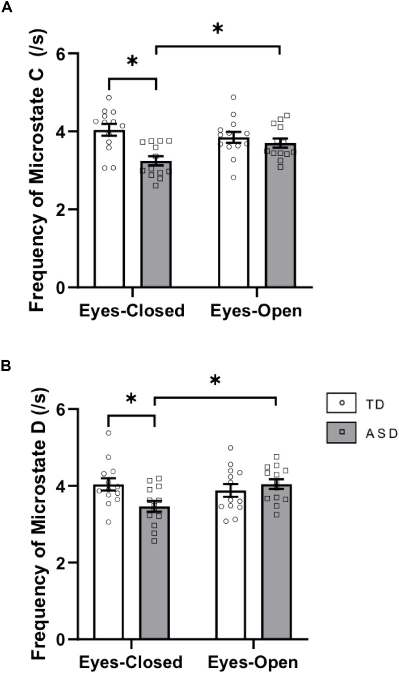
Although fMRI studies have produced considerable evidence for differences in the spatial connectivity of resting-state brain networks in persons with autism spectrum disorder (ASD) relative to typically developing (TD) peers, little is known about the temporal dynamics of these brain networks in ASD. The aim of this study was to examine the EEG microstate architecture in children with ASD as compared to TD at rest in two separate conditions - eyes-closed (EC) and eyes-open (EO). EEG microstate analysis was performed on resting-state data of 13 ASD and 13 TD children matched on age, gender, and IQ. We found that children with ASD and TD peers produced topographically similar canonical microstates at rest. Group differences in the duration and frequency of these microstates were found primarily in the EC resting-state condition. In line with previous fMRI findings that have reported differences in spatial connectivity within the salience network (previously correlated with the activity of microstate C) in ASD, we found that the duration of activation of microstate C was increased, and the frequency of microstate C was decreased in ASD as compared to TD in EC resting-state. Functionally, these results may be reflective of alterations in interoceptive processes in ASD. These results suggest a unique pattern of EEG microstate architecture in ASD relative to TD during resting-states and also that EEG microstate parameters in ASD are susceptible to differences in resting-state conditions.
尽管功能磁共振成像(fMRI)研究已经产生了大量证据,表明自闭症谱系障碍(ASD)患者相对于正常发育(TD)的同龄人,其静息态脑网络的空间连接存在差异,但对于ASD中这些脑网络的时间动态却知之甚少。本研究的目的是在两种不同的条件下——闭眼(EC)和睁眼(EO),比较ASD儿童与TD儿童静息时的脑电图微状态结构。对13名ASD儿童和13名在年龄、性别和智商上匹配的TD儿童的静息态数据进行了脑电图微状态分析。我们发现,ASD儿童和TD同龄人在静息时产生的典型微状态在地形上相似。这些微状态的持续时间和频率的组间差异主要出现在EC静息状态下。与之前的fMRI研究结果一致,该研究报告了ASD中突显网络内空间连接的差异(之前与微状态C的活动相关),我们发现,在EC静息状态下,与TD相比,ASD中微状态C的激活持续时间增加,微状态C的频率降低。在功能上,这些结果可能反映了ASD中内感受过程的改变。这些结果表明,与TD相比,ASD在静息状态下具有独特的脑电图微状态结构模式,并且ASD中的脑电图微状态参数易受静息状态条件差异的影响。