• 文献检索
  • 文档翻译
  • 深度研究
  • 学术资讯
  • Suppr Zotero 插件Zotero 插件
  • 邀请有礼
  • 套餐&价格
  • 历史记录
应用&插件
Suppr Zotero 插件Zotero 插件浏览器插件Mac 客户端Windows 客户端微信小程序
定价
高级版会员购买积分包购买API积分包
服务
文献检索文档翻译深度研究API 文档MCP 服务
关于我们
关于 Suppr公司介绍联系我们用户协议隐私条款
关注我们

Suppr 超能文献

核心技术专利:CN118964589B侵权必究
粤ICP备2023148730 号-1Suppr @ 2026

文献检索

告别复杂PubMed语法,用中文像聊天一样搜索,搜遍4000万医学文献。AI智能推荐,让科研检索更轻松。

立即免费搜索

文件翻译

保留排版,准确专业,支持PDF/Word/PPT等文件格式,支持 12+语言互译。

免费翻译文档

深度研究

AI帮你快速写综述,25分钟生成高质量综述,智能提取关键信息,辅助科研写作。

立即免费体验

磁共振成像用于评估同种异体肾移植中的间质纤维化

Magnetic Resonance Imaging for Evaluation of Interstitial Fibrosis in Kidney Allografts.

作者信息

Beck-Tölly Andrea, Eder Michael, Beitzke Dietrich, Eskandary Farsad, Agibetov Asan, Lampichler Katharina, Hamböck Martina, Regele Heinz, Kläger Johannes, Nackenhorst Maja, Böhmig Georg A

机构信息

Division of Cardiovascular and Interventional Radiology, Department of Biomedical Imaging and Image-Guided Therapy, Medical University of Vienna, Vienna, Austria.

Division of Nephrology and Dialysis, Department of Medicine III, Medical University of Vienna, Vienna, Austria.

出版信息

Transplant Direct. 2020 Jul 15;6(8):e577. doi: 10.1097/TXD.0000000000001009. eCollection 2020 Aug.

DOI:10.1097/TXD.0000000000001009
PMID:33134501
原文链接:https://pmc.ncbi.nlm.nih.gov/articles/PMC7581173/
Abstract

UNLABELLED

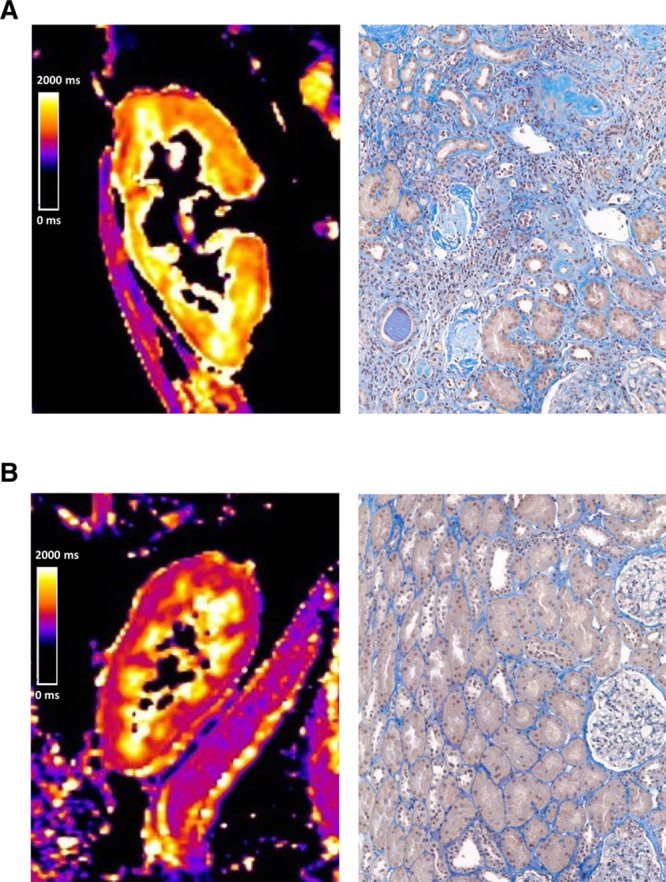
Interstitial fibrosis (IF) is the common pathway of chronic kidney injury in various conditions. Magnetic resonance imaging (MRI) may be a promising tool for the noninvasive assessment of IF in renal allografts.

METHODS

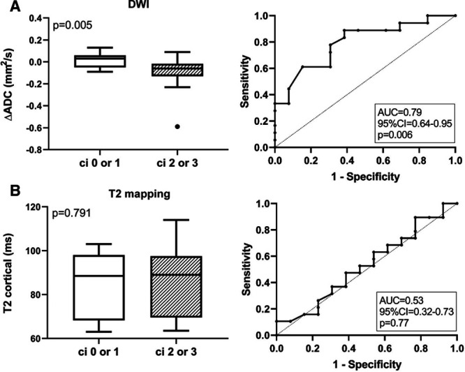
This prospective trial was primarily designed to investigate whether the results of T1-weighted MRI associate with the degree of IF. Thirty-two kidney transplant recipients were subjected to 1.5-Tesla MRI scans shortly before or after routine allograft biopsies. MRI parameters [T1 and T2 relaxation times; apparent diffusion coefficient (ADC)] were assessed for cortical and medullary sections.

RESULTS

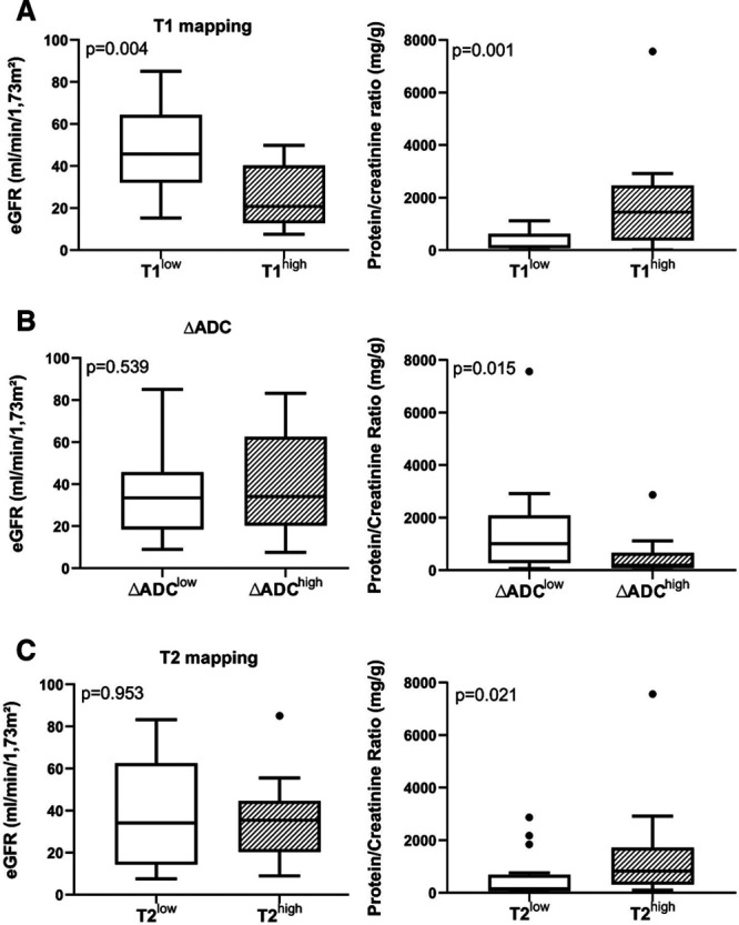
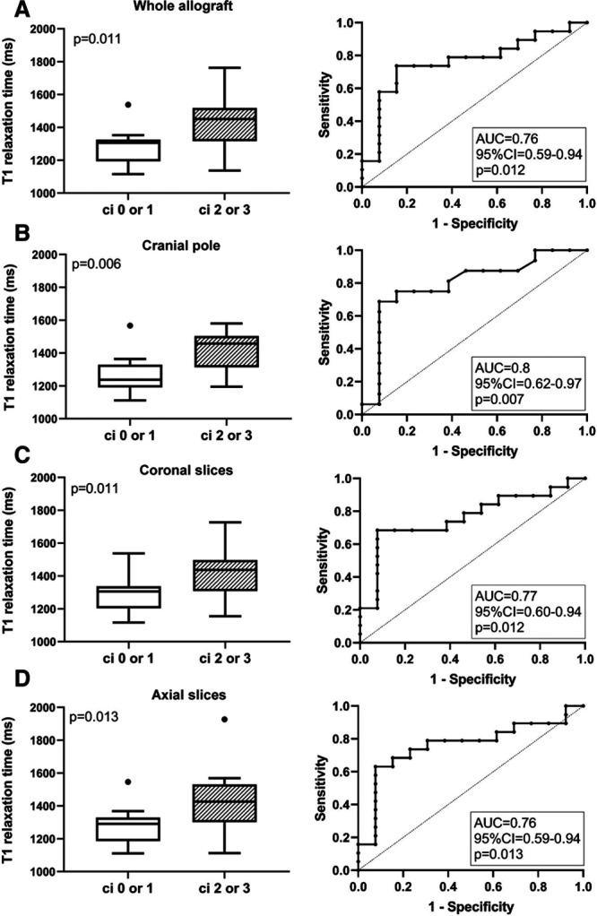
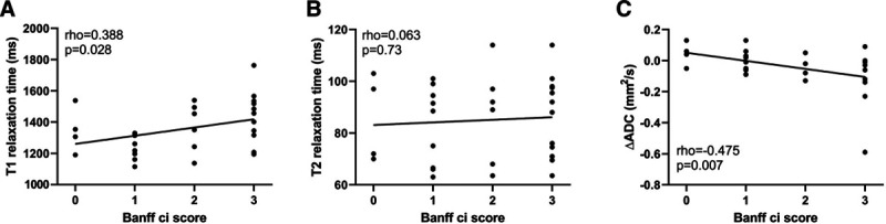
Advanced IF (Banff ci score >1) was associated with higher cortical T1 (but not T2) values [1451 (median; interquartile range: 1331-1506) versus 1306 (1197-1321) ms in subjects with ci scores ≤1; = 0.011; receiver operating characteristic area under the curve for prediction of ci > 1: 0.76]. In parallel, T1 values were associated with kidney function and proteinuria. There was also a relationship between IF and corticomedullary differences on ADC maps (receiver operating characteristic area under the curve for prediction of ci ≤ 1: 0.79).

CONCLUSIONS

Our results support the use of MRI for noninvasive assessment of allograft scarring. Future studies will have to clarify the role of T1 (and ADC) mapping as a surrogate endpoint reflecting the progression of chronic graft damage.

摘要

未标注

间质纤维化(IF)是各种情况下慢性肾损伤的共同途径。磁共振成像(MRI)可能是一种用于无创评估肾移植中IF的有前景的工具。

方法

这项前瞻性试验主要旨在研究T1加权MRI结果是否与IF程度相关。32名肾移植受者在常规移植肾活检前后不久接受了1.5特斯拉MRI扫描。对皮质和髓质部分评估MRI参数[T1和T2弛豫时间;表观扩散系数(ADC)]。

结果

高级IF(Banff ci评分>1)与较高的皮质T1(而非T2)值相关[ci评分≤1的受试者中为1451(中位数;四分位间距:1331 - 1506)对1306(1197 - 1321)毫秒;P = 0.011;预测ci>1的曲线下受试者工作特征面积:0.76]。同时,T1值与肾功能和蛋白尿相关。IF与ADC图上的皮质髓质差异之间也存在关系(预测ci≤1的曲线下受试者工作特征面积:0.79)。

结论

我们的结果支持使用MRI对移植肾瘢痕进行无创评估。未来的研究将必须阐明T1(和ADC)成像作为反映慢性移植肾损伤进展的替代终点的作用。

https://cdn.ncbi.nlm.nih.gov/pmc/blobs/e5b8/7581173/7f7402768c8d/txd-6-e577-g009.jpg
https://cdn.ncbi.nlm.nih.gov/pmc/blobs/e5b8/7581173/b819baf94309/txd-6-e577-g004.jpg
https://cdn.ncbi.nlm.nih.gov/pmc/blobs/e5b8/7581173/0d923ecd2f20/txd-6-e577-g005.jpg
https://cdn.ncbi.nlm.nih.gov/pmc/blobs/e5b8/7581173/a766e2011d19/txd-6-e577-g007.jpg
https://cdn.ncbi.nlm.nih.gov/pmc/blobs/e5b8/7581173/a4d8cbc0645b/txd-6-e577-g008.jpg
https://cdn.ncbi.nlm.nih.gov/pmc/blobs/e5b8/7581173/7f7402768c8d/txd-6-e577-g009.jpg

相似文献

1
Magnetic Resonance Imaging for Evaluation of Interstitial Fibrosis in Kidney Allografts.磁共振成像用于评估同种异体肾移植中的间质纤维化
Transplant Direct. 2020 Jul 15;6(8):e577. doi: 10.1097/TXD.0000000000001009. eCollection 2020 Aug.
2
Combination of Functional Magnetic Resonance Imaging and Histopathologic Analysis to Evaluate Interstitial Fibrosis in Kidney Allografts.功能磁共振成像与组织病理学分析联合评估肾移植间质纤维化。
Clin J Am Soc Nephrol. 2019 Sep 6;14(9):1372-1380. doi: 10.2215/CJN.00020119. Epub 2019 Aug 15.
3
Multiparametric magnetic resonance imaging shows promising results to assess renal transplant dysfunction with fibrosis.多参数磁共振成像在评估伴有纤维化的肾移植功能障碍方面显示出有希望的结果。
Kidney Int. 2020 Feb;97(2):414-420. doi: 10.1016/j.kint.2019.09.030. Epub 2019 Oct 30.
4
Evaluation of interstitial fibrosis in chronic kidney disease by multiparametric functional MRI and histopathologic analysis.多参数功能磁共振成像与组织病理学分析评估慢性肾脏病间质纤维化。
Eur Radiol. 2023 Jun;33(6):4138-4147. doi: 10.1007/s00330-022-09329-7. Epub 2022 Dec 11.
5
Validation of the corticomedullary difference in magnetic resonance imaging-derived apparent diffusion coefficient for kidney fibrosis detection: a cross-sectional study.基于磁共振成像表观扩散系数的皮质-髓质差异对肾脏纤维化检测的验证:一项横断面研究。
Nephrol Dial Transplant. 2020 Jun 1;35(6):937-945. doi: 10.1093/ndt/gfy389.
6
Diffusion-Weighted Imaging and Mapping of T1 and T2 Relaxation Time for Evaluation of Chronic Renal Allograft Rejection in a Translational Mouse Model.在一个转化小鼠模型中,利用扩散加权成像以及T1和T2弛豫时间映射来评估慢性同种异体肾移植排斥反应
J Clin Med. 2021 Sep 23;10(19):4318. doi: 10.3390/jcm10194318.
7
Multiparametric Assessment of Changes in Renal Tissue after Kidney Transplantation with Quantitative MR Relaxometry and Diffusion-Tensor Imaging at 3 T.3T磁共振定量弛豫测量法和扩散张量成像对肾移植后肾组织变化的多参数评估
J Clin Med. 2020 May 21;9(5):1551. doi: 10.3390/jcm9051551.
8
Quantitative assessment of renal structural and functional changes in chronic kidney disease using multi-parametric magnetic resonance imaging.使用多参数磁共振成像技术定量评估慢性肾脏病的肾脏结构和功能变化。
Nephrol Dial Transplant. 2020 Jun 1;35(6):955-964. doi: 10.1093/ndt/gfz129.
9
T2 relaxation time and apparent diffusion coefficient for noninvasive assessment of renal pathology after acute kidney injury in mice: comparison with histopathology.T2 弛豫时间和表观扩散系数在急性肾损伤后小鼠肾脏病变的无创评估中的应用:与组织病理学的比较。
Invest Radiol. 2013 Dec;48(12):834-42. doi: 10.1097/RLI.0b013e31829d0414.
10
Arterial spin labeling combined with T1 mapping for assessment of kidney function and histopathology in patients with long-term renal transplant survival after kidney transplantation.动脉自旋标记结合T1映射用于评估肾移植后长期肾移植存活患者的肾功能和组织病理学。
Quant Imaging Med Surg. 2024 Mar 15;14(3):2415-2425. doi: 10.21037/qims-23-1577. Epub 2024 Mar 4.

引用本文的文献

1
The Possibilities of Multiparametric Magnetic Resonance Imaging to Reflect Functional and Structural Graft Changes 1 Year After Kidney Transplantation.多参数磁共振成像反映肾移植术后1年移植物功能和结构变化的可能性
Medicina (Kaunas). 2025 Jul 13;61(7):1268. doi: 10.3390/medicina61071268.
2
Prediction of early renal allograft dysfunction using multiparametric magnetic resonance imaging.使用多参数磁共振成像预测早期肾移植功能障碍
Quant Imaging Med Surg. 2025 May 1;15(5):4333-4342. doi: 10.21037/qims-24-2182. Epub 2025 Apr 22.
3
T Relaxation Time for the Prediction of Renal Transplant Dysfunction.

本文引用的文献

1
Myocardial Tissue Characterization and Fibrosis by Imaging.心肌组织的影像学特征与纤维化
JACC Cardiovasc Imaging. 2020 May;13(5):1221-1234. doi: 10.1016/j.jcmg.2019.06.030. Epub 2019 Sep 18.
2
Combination of Functional Magnetic Resonance Imaging and Histopathologic Analysis to Evaluate Interstitial Fibrosis in Kidney Allografts.功能磁共振成像与组织病理学分析联合评估肾移植间质纤维化。
Clin J Am Soc Nephrol. 2019 Sep 6;14(9):1372-1380. doi: 10.2215/CJN.00020119. Epub 2019 Aug 15.
3
Validation of the corticomedullary difference in magnetic resonance imaging-derived apparent diffusion coefficient for kidney fibrosis detection: a cross-sectional study.
用于预测肾移植功能障碍的T弛豫时间。
Transpl Int. 2025 Apr 10;38:14301. doi: 10.3389/ti.2025.14301. eCollection 2025.
4
T1 mapping magnetic resonance imaging predicts decline of kidney function.T1 映射磁共振成像可预测肾功能衰退。
Clin Kidney J. 2025 Feb 7;18(3):sfaf032. doi: 10.1093/ckj/sfaf032. eCollection 2025 Mar.
5
Potential MRI Biomarkers for Predicting Kidney Function and Histological Damage in Transplanted Deceased Donor Kidney Recipients.预测已故供体肾移植受者肾功能和组织学损伤的潜在MRI生物标志物
J Clin Med. 2025 Feb 18;14(4):1349. doi: 10.3390/jcm14041349.
6
Histopathological correlations of CT-based radiomics imaging biomarkers in native kidney biopsy.基于 CT 的放射组学成像生物标志物在原发性肾脏活检中的组织病理学相关性。
BMC Med Imaging. 2024 Sep 27;24(1):256. doi: 10.1186/s12880-024-01434-x.
7
Magnetic resonance elastography resolving all gross anatomical segments of the kidney during controlled hydration.在控制性水化过程中对肾脏所有大体解剖节段进行分辨的磁共振弹性成像。
Front Physiol. 2024 Feb 7;15:1327407. doi: 10.3389/fphys.2024.1327407. eCollection 2024.
8
Magnetic Resonance Imaging in Clinical Trials of Diabetic Kidney Disease.糖尿病肾病临床试验中的磁共振成像
J Clin Med. 2023 Jul 11;12(14):4625. doi: 10.3390/jcm12144625.
9
Contemporary and Emerging MRI Strategies for Assessing Kidney Allograft Complications: Arterial Stenosis and Parenchymal Injury, From the Special Series on Imaging of Fibrosis.评估肾移植并发症的现代和新兴 MRI 策略:动脉狭窄和实质损伤,来自纤维化成像特刊。
AJR Am J Roentgenol. 2024 Mar;222(3):e2329418. doi: 10.2214/AJR.23.29418. Epub 2023 Jun 14.
10
Renal MRI: From Nephron to NMR Signal.肾脏 MRI:从肾单位到 NMR 信号。
J Magn Reson Imaging. 2023 Dec;58(6):1660-1679. doi: 10.1002/jmri.28828. Epub 2023 May 26.
基于磁共振成像表观扩散系数的皮质-髓质差异对肾脏纤维化检测的验证:一项横断面研究。
Nephrol Dial Transplant. 2020 Jun 1;35(6):937-945. doi: 10.1093/ndt/gfy389.
4
Magnetic resonance imaging T1- and T2-mapping to assess renal structure and function: a systematic review and statement paper.磁共振成像 T1 和 T2 映射评估肾脏结构和功能:系统评价和立场声明。
Nephrol Dial Transplant. 2018 Sep 1;33(suppl_2):ii41-ii50. doi: 10.1093/ndt/gfy198.
5
A 2018 Reference Guide to the Banff Classification of Renal Allograft Pathology.2018 年肾移植病理的班夫分类参考指南。
Transplantation. 2018 Nov;102(11):1795-1814. doi: 10.1097/TP.0000000000002366.
6
The Prognostic Value of Histopathologic Lesions in Native Kidney Biopsy Specimens: Results from the Boston Kidney Biopsy Cohort Study.原发性肾脏活检组织学病变的预后价值:来自波士顿肾脏活检队列研究的结果。
J Am Soc Nephrol. 2018 Aug;29(8):2213-2224. doi: 10.1681/ASN.2017121260. Epub 2018 Jun 4.
7
The Banff 2017 Kidney Meeting Report: Revised diagnostic criteria for chronic active T cell-mediated rejection, antibody-mediated rejection, and prospects for integrative endpoints for next-generation clinical trials.Banff 2017 年会肾脏报告:慢性活动性 T 细胞介导排斥反应、抗体介导排斥反应的修订诊断标准,以及下一代临床试验综合终点的前景。
Am J Transplant. 2018 Feb;18(2):293-307. doi: 10.1111/ajt.14625. Epub 2018 Jan 21.
8
Magnetic Resonance Elastography to Assess Fibrosis in Kidney Allografts.磁共振弹性成像评估肾移植中的纤维化。
Clin J Am Soc Nephrol. 2017 Oct 6;12(10):1671-1679. doi: 10.2215/CJN.01830217. Epub 2017 Aug 30.
9
Assessment of acute kidney injury with T1 mapping MRI following solid organ transplantation.评估实体器官移植后 T1 mapping MRI 检查的急性肾损伤。
Eur Radiol. 2018 Jan;28(1):44-50. doi: 10.1007/s00330-017-4943-4. Epub 2017 Jul 14.
10
Kidney Fibrosis: Origins and Interventions.肾纤维化:起源与干预措施
Transplantation. 2017 Apr;101(4):713-726. doi: 10.1097/TP.0000000000001608.