• 文献检索
  • 文档翻译
  • 深度研究
  • 学术资讯
  • Suppr Zotero 插件Zotero 插件
  • 邀请有礼
  • 套餐&价格
  • 历史记录
应用&插件
Suppr Zotero 插件Zotero 插件浏览器插件Mac 客户端Windows 客户端微信小程序
定价
高级版会员购买积分包购买API积分包
服务
文献检索文档翻译深度研究API 文档MCP 服务
关于我们
关于 Suppr公司介绍联系我们用户协议隐私条款
关注我们

Suppr 超能文献

核心技术专利:CN118964589B侵权必究
粤ICP备2023148730 号-1Suppr @ 2026

文献检索

告别复杂PubMed语法,用中文像聊天一样搜索,搜遍4000万医学文献。AI智能推荐,让科研检索更轻松。

立即免费搜索

文件翻译

保留排版,准确专业,支持PDF/Word/PPT等文件格式,支持 12+语言互译。

免费翻译文档

深度研究

AI帮你快速写综述,25分钟生成高质量综述,智能提取关键信息,辅助科研写作。

立即免费体验

单细胞分辨率解析脑动静脉畸形中内皮细胞的多样性。

Mapping endothelial-cell diversity in cerebral cavernous malformations at single-cell resolution.

机构信息

Vascular Biology Unit, FIRC Institute of Molecular Oncology Foundation (IFOM), Milan, Italy.

Department of Immunology, Genetics and Pathology, Uppsala University, Uppsala, Sweden.

出版信息

Elife. 2020 Nov 3;9:e61413. doi: 10.7554/eLife.61413.

DOI:10.7554/eLife.61413
PMID:33138917
原文链接:https://pmc.ncbi.nlm.nih.gov/articles/PMC7609066/
Abstract

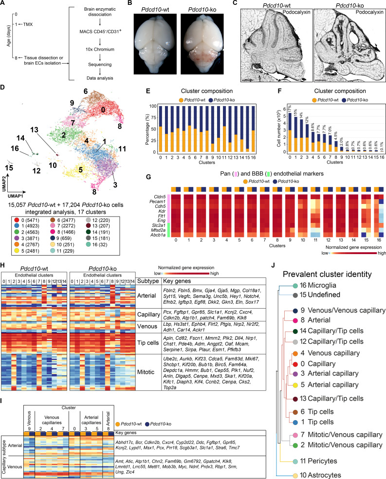
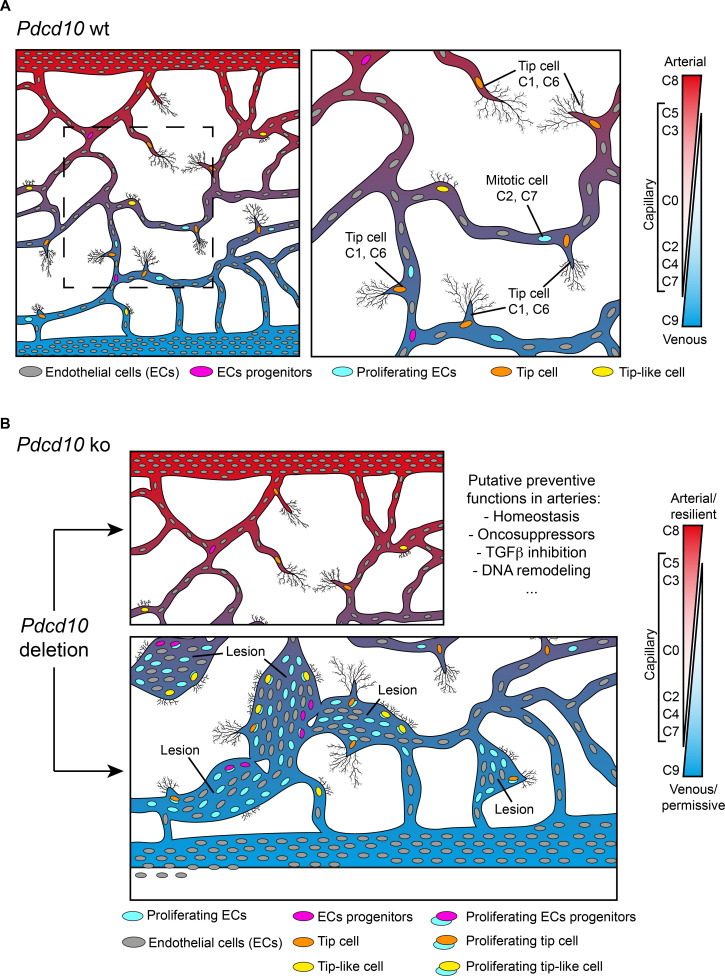
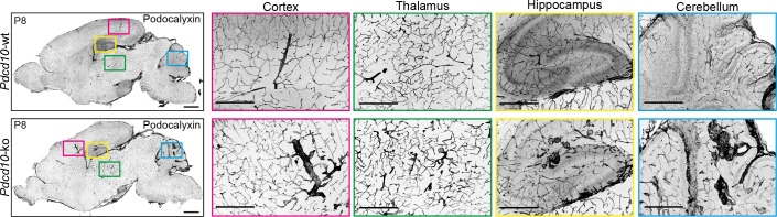
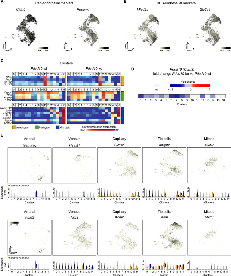
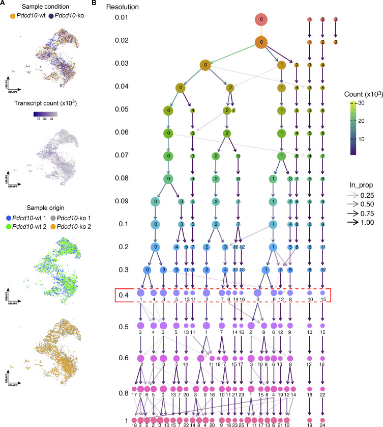
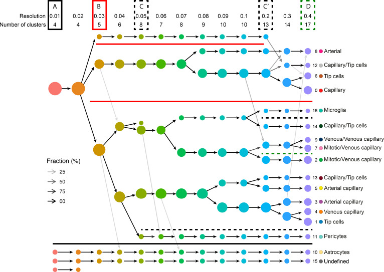
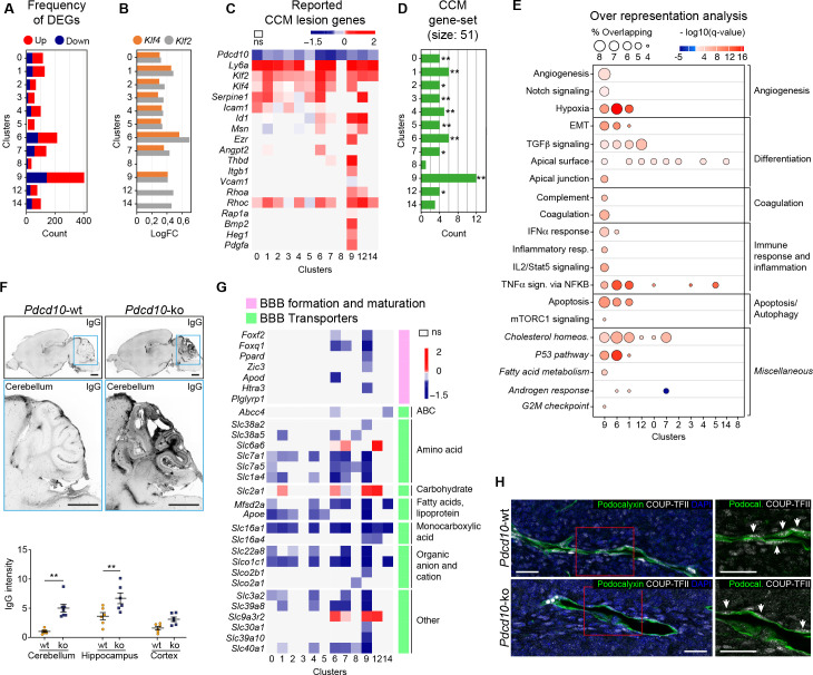
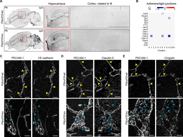
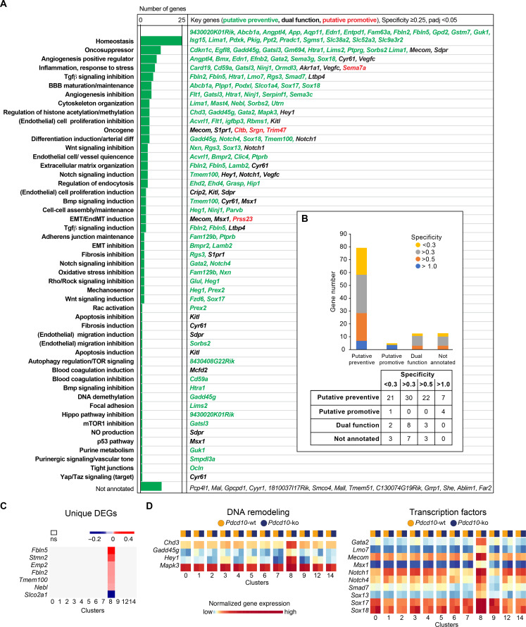
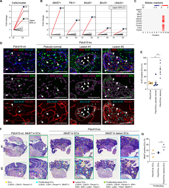
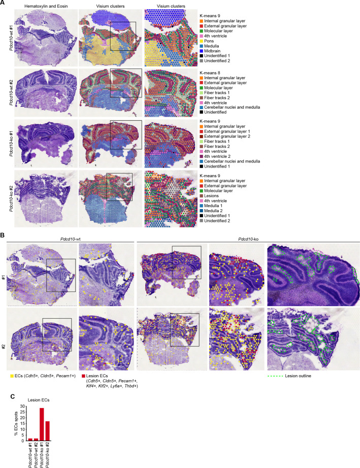
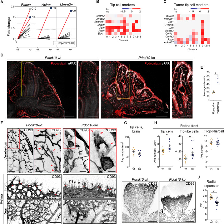
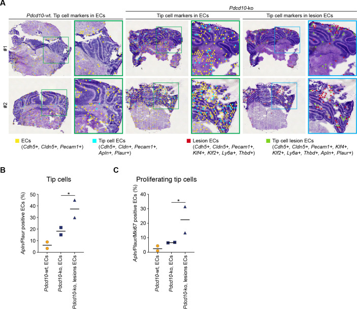
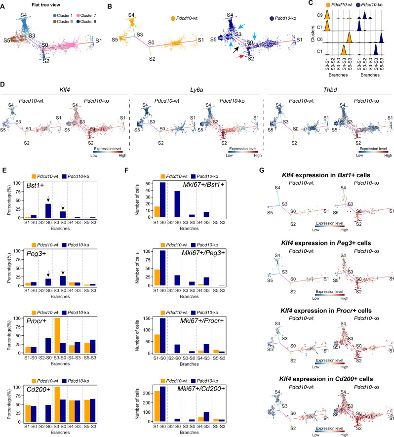
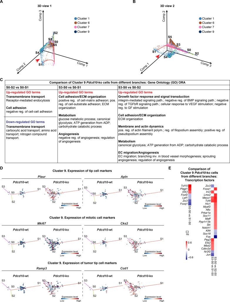
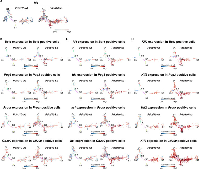
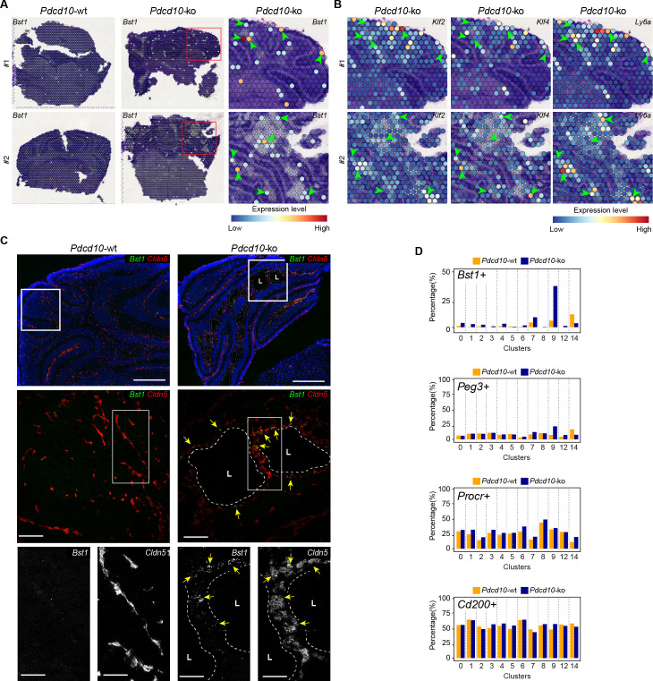
Cerebral cavernous malformation (CCM) is a rare neurovascular disease that is characterized by enlarged and irregular blood vessels that often lead to cerebral hemorrhage. Loss-of-function mutations to any of three genes results in CCM lesion formation; namely, , , and . Here, we report for the first time in-depth single-cell RNA sequencing, combined with spatial transcriptomics and immunohistochemistry, to comprehensively characterize subclasses of brain endothelial cells (ECs) under both normal conditions and after deletion of ( in a mouse model of CCM. Integrated single-cell analysis identifies arterial ECs as refractory to CCM transformation. Conversely, a subset of angiogenic venous capillary ECs and respective resident endothelial progenitors appear to be at the origin of CCM lesions. These data are relevant for the understanding of the plasticity of the brain vascular system and provide novel insights into the molecular basis of CCM disease at the single cell level.

摘要

脑 腔 隙 性 动 脉 瘤(CCM)是 一 种 罕 见 的 神 经 血 管 疾 病,其 特 征 是 血 管 扩 张 和 不 规 则,这 常 导 致 脑 出 血。三 种 基 因 的 功 能 失 活 突 变 导 致 CCM 病 灶 的 形 成;即 、 和 。在 这 里,我 们 首 次 报 道 了 深 度 的 单 细 胞 RNA 测 序,结 合 空 间 转 录 组 学 和 免 疫 组 化,全 面 描 述 了 在 正 常 情 况 下 和 小 鼠 CCM 模 型 中 (基 因 缺 失 后 的 脑 内 皮 细 胞(ECs)的 亚 类。集 成 的 单 细 胞 分 析 确 认 动 脉 ECs 对 CCM 转 化 具 有 抵 抗 性。相 反,一 部 分 成 血 管 的 静 脉 毛 细 血 管 ECs 和 相 应 的 内 皮 祖 细 胞 看 来 是 CCM 病 灶 的 起 源。这 些 数 据 对 于 理 解 脑 血 管 系 统 的 塑 性 具 有 重 要 意 义,并 为 单 细 胞 水 平 的 CCM 疾 病 的 分 子 基 础 提 供 了 新 的 见 解。

https://cdn.ncbi.nlm.nih.gov/pmc/blobs/544c/7609066/63843b0b958a/elife-61413-fig8.jpg
https://cdn.ncbi.nlm.nih.gov/pmc/blobs/544c/7609066/f6478637b79d/elife-61413-fig1.jpg
https://cdn.ncbi.nlm.nih.gov/pmc/blobs/544c/7609066/7b9cb001b805/elife-61413-fig1-figsupp1.jpg
https://cdn.ncbi.nlm.nih.gov/pmc/blobs/544c/7609066/14ca4cb771d3/elife-61413-fig1-figsupp2.jpg
https://cdn.ncbi.nlm.nih.gov/pmc/blobs/544c/7609066/e6ffd049a9db/elife-61413-fig1-figsupp3.jpg
https://cdn.ncbi.nlm.nih.gov/pmc/blobs/544c/7609066/0f28f357e5b7/elife-61413-fig1-figsupp4.jpg
https://cdn.ncbi.nlm.nih.gov/pmc/blobs/544c/7609066/93036abc4ccd/elife-61413-fig2.jpg
https://cdn.ncbi.nlm.nih.gov/pmc/blobs/544c/7609066/f571c6bbd882/elife-61413-fig2-figsupp1.jpg
https://cdn.ncbi.nlm.nih.gov/pmc/blobs/544c/7609066/df65d50255d0/elife-61413-fig3.jpg
https://cdn.ncbi.nlm.nih.gov/pmc/blobs/544c/7609066/4af336dba7d0/elife-61413-fig4.jpg
https://cdn.ncbi.nlm.nih.gov/pmc/blobs/544c/7609066/102dff9a14ca/elife-61413-fig4-figsupp1.jpg
https://cdn.ncbi.nlm.nih.gov/pmc/blobs/544c/7609066/2bfe745f614f/elife-61413-fig5.jpg
https://cdn.ncbi.nlm.nih.gov/pmc/blobs/544c/7609066/850e980085fd/elife-61413-fig5-figsupp1.jpg
https://cdn.ncbi.nlm.nih.gov/pmc/blobs/544c/7609066/91145aaab0c0/elife-61413-fig6.jpg
https://cdn.ncbi.nlm.nih.gov/pmc/blobs/544c/7609066/4a68e638b192/elife-61413-fig6-figsupp1.jpg
https://cdn.ncbi.nlm.nih.gov/pmc/blobs/544c/7609066/1208df6cffcf/elife-61413-fig6-figsupp2.jpg
https://cdn.ncbi.nlm.nih.gov/pmc/blobs/544c/7609066/5f39d82008df/elife-61413-fig7.jpg
https://cdn.ncbi.nlm.nih.gov/pmc/blobs/544c/7609066/63843b0b958a/elife-61413-fig8.jpg

相似文献

1
Mapping endothelial-cell diversity in cerebral cavernous malformations at single-cell resolution.单细胞分辨率解析脑动静脉畸形中内皮细胞的多样性。
Elife. 2020 Nov 3;9:e61413. doi: 10.7554/eLife.61413.
2
mTORC1 Signaling in Brain Endothelial Progenitors Contributes to CCM Pathogenesis.mTORC1 信号在脑内皮祖细胞中的作用促进 CCM 发病机制。
Circ Res. 2024 Aug 2;135(4):e94-e113. doi: 10.1161/CIRCRESAHA.123.324015. Epub 2024 Jul 3.
3
Differential angiogenesis function of CCM2 and CCM3 in cerebral cavernous malformations.CCM2 和 CCM3 在脑动静脉畸形中的差异血管生成功能。
Neurosurg Focus. 2010 Sep;29(3):E1. doi: 10.3171/2010.5.FOCUS1090.
4
Mural Cell-Specific Deletion of Cerebral Cavernous Malformation 3 in the Brain Induces Cerebral Cavernous Malformations.脑内血管性腔隙畸形 3 的壁细胞特异性缺失可诱导脑内血管性腔隙畸形。
Arterioscler Thromb Vasc Biol. 2020 Sep;40(9):2171-2186. doi: 10.1161/ATVBAHA.120.314586. Epub 2020 Jul 9.
5
Caveolae-mediated Tie2 signaling contributes to CCM pathogenesis in a brain endothelial cell-specific Pdcd10-deficient mouse model.小窝介导的 Tie2 信号通路促进了脑内皮细胞特异性 Pdcd10 缺失小鼠模型中 CCM 的发病机制。
Nat Commun. 2021 Jan 25;12(1):504. doi: 10.1038/s41467-020-20774-0.
6
Conditional deletion of Ccm2 causes hemorrhage in the adult brain: a mouse model of human cerebral cavernous malformations.条件性敲除 Ccm2 可导致成年大脑出血:人类脑海绵状血管畸形的小鼠模型。
Hum Mol Genet. 2011 Aug 15;20(16):3198-206. doi: 10.1093/hmg/ddr225. Epub 2011 May 19.
7
CCM3 and cerebral cavernous malformation disease.CCM3 与脑静脉血管畸形病。
Stroke Vasc Neurol. 2019 Mar 2;4(2):67-70. doi: 10.1136/svn-2018-000195. eCollection 2019 Jul.
8
Neuroinflammation Plays a Critical Role in Cerebral Cavernous Malformation Disease.神经炎症在脑动静脉畸形疾病中发挥着关键作用。
Circ Res. 2022 Nov 11;131(11):909-925. doi: 10.1161/CIRCRESAHA.122.321129. Epub 2022 Oct 26.
9
Distinct cellular roles for PDCD10 define a gut-brain axis in cerebral cavernous malformation.PDCD10 在不同细胞中的作用定义了脑动静脉畸形的肠脑轴。
Sci Transl Med. 2019 Nov 27;11(520). doi: 10.1126/scitranslmed.aaw3521.
10
Astrocytes propel neurovascular dysfunction during cerebral cavernous malformation lesion formation.星形胶质细胞在脑海绵状血管畸形病变形成过程中推动神经血管功能障碍。
J Clin Invest. 2021 Jul 1;131(13). doi: 10.1172/JCI139570.

引用本文的文献

1
High-throughput differentiation of human blood vessel organoids reveals overlapping and distinct functions of the cerebral cavernous malformation proteins.人血管类器官的高通量分化揭示了脑海绵状血管畸形蛋白的重叠和独特功能。
Angiogenesis. 2025 Jun 6;28(3):32. doi: 10.1007/s10456-025-09985-5.
2
Embryological cellular origins and hypoxia-mediated mechanisms in PIK3CA-driven refractory vascular malformations.PIK3CA驱动的难治性血管畸形的胚胎细胞起源及缺氧介导机制
EMBO Mol Med. 2025 Apr 16. doi: 10.1038/s44321-025-00235-1.
3
Mapping the transcriptional and epigenetic landscape of organotypic endothelial diversity in the developing and adult mouse.

本文引用的文献

1
Blocking Signalopathic Events to Treat Cerebral Cavernous Malformations.阻断信号病变事件以治疗脑海绵状血管畸形
Trends Mol Med. 2020 Sep;26(9):874-887. doi: 10.1016/j.molmed.2020.03.003. Epub 2020 Apr 3.
2
An Integrated Gene Expression Landscape Profiling Approach to Identify Lung Tumor Endothelial Cell Heterogeneity and Angiogenic Candidates.一种用于识别肺肿瘤内皮细胞异质性和血管生成候选物的综合基因表达谱分析方法。
Cancer Cell. 2020 Mar 16;37(3):421. doi: 10.1016/j.ccell.2020.03.002.
3
Single-Cell Transcriptome Atlas of Murine Endothelial Cells.
绘制发育中和成年小鼠器官型内皮多样性的转录和表观遗传图谱。
Nat Cardiovasc Res. 2025 Apr;4(4):473-495. doi: 10.1038/s44161-025-00618-0. Epub 2025 Mar 17.
4
Single-Cell Analysis in Cerebrovascular Research: Primed for Breakthroughs and Clinical Impact.脑血管研究中的单细胞分析:有望取得突破并产生临床影响。
Stroke. 2025 Apr;56(4):1082-1091. doi: 10.1161/STROKEAHA.124.049001. Epub 2024 Dec 30.
5
Pharmacological blocking of neutrophil extracellular traps attenuates immunothrombosis and neuroinflammation in cerebral cavernous malformation.中性粒细胞胞外诱捕网的药理学阻断可减轻海绵状血管瘤中的免疫血栓形成和神经炎症。
Nat Cardiovasc Res. 2024 Dec;3(12):1549-1567. doi: 10.1038/s44161-024-00577-y. Epub 2024 Dec 4.
6
Force-mediated recruitment and reprogramming of healthy endothelial cells drive vascular lesion growth.力介导的健康内皮细胞募集和重编程驱动血管病变生长。
Nat Commun. 2024 Oct 6;15(1):8660. doi: 10.1038/s41467-024-52866-6.
7
When, where and which PIK3CA mutations are pathogenic in congenital disorders.PIK3CA基因的哪些突变在先天性疾病中具有致病性,以及这些突变发生的时间和位置。
Nat Cardiovasc Res. 2022 Aug;1(8):700-714. doi: 10.1038/s44161-022-00107-8. Epub 2022 Aug 8.
8
Dynamics of Endothelial Cell Diversity and Plasticity in Health and Disease.内皮细胞多样性和可塑性在健康和疾病中的动态变化。
Cells. 2024 Jul 29;13(15):1276. doi: 10.3390/cells13151276.
9
Single-cell atlas of the human brain vasculature across development, adulthood and disease.人类大脑血管系统在发育、成年和疾病过程中的单细胞图谱
Nature. 2024 Aug;632(8025):603-613. doi: 10.1038/s41586-024-07493-y. Epub 2024 Jul 10.
10
mTORC1 Signaling in Brain Endothelial Progenitors Contributes to CCM Pathogenesis.mTORC1 信号在脑内皮祖细胞中的作用促进 CCM 发病机制。
Circ Res. 2024 Aug 2;135(4):e94-e113. doi: 10.1161/CIRCRESAHA.123.324015. Epub 2024 Jul 3.
单细胞转录组图谱:鼠类血管内皮细胞
Cell. 2020 Feb 20;180(4):764-779.e20. doi: 10.1016/j.cell.2020.01.015. Epub 2020 Feb 13.
4
Low fluid shear stress conditions contribute to activation of cerebral cavernous malformation signalling pathways.低流体切应力条件有助于激活脑海绵状血管畸形信号通路。
Biochim Biophys Acta Mol Basis Dis. 2019 Nov 1;1865(11):165519. doi: 10.1016/j.bbadis.2019.07.013. Epub 2019 Jul 29.
5
Endothelial cell clonal expansion in the development of cerebral cavernous malformations.脑动静脉畸形发生中内皮细胞的克隆性扩张。
Nat Commun. 2019 Jun 24;10(1):2761. doi: 10.1038/s41467-019-10707-x.
6
Comprehensive Integration of Single-Cell Data.单细胞数据的综合整合。
Cell. 2019 Jun 13;177(7):1888-1902.e21. doi: 10.1016/j.cell.2019.05.031. Epub 2019 Jun 6.
7
Single-cell trajectories reconstruction, exploration and mapping of omics data with STREAM.使用 STREAM 进行单细胞轨迹重建、组学数据探索和映射。
Nat Commun. 2019 Apr 23;10(1):1903. doi: 10.1038/s41467-019-09670-4.
8
CDC42 Deletion Elicits Cerebral Vascular Malformations via Increased MEKK3-Dependent KLF4 Expression.CDC42 缺失通过增加 MEKK3 依赖性 KLF4 表达引发脑血管畸形。
Circ Res. 2019 Apr 12;124(8):1240-1252. doi: 10.1161/CIRCRESAHA.118.314300.
9
Cerebral cavernous malformations form an anticoagulant vascular domain in humans and mice.脑内海绵状血管畸形在人类和小鼠中形成一个抗凝血的血管区域。
Blood. 2019 Jan 17;133(3):193-204. doi: 10.1182/blood-2018-06-856062. Epub 2018 Nov 15.
10
Transcriptional and epigenomic landscapes of CNS and non-CNS vascular endothelial cells.中枢神经系统和非中枢神经系统血管内皮细胞的转录组和表观基因组图谱。
Elife. 2018 Sep 6;7:e36187. doi: 10.7554/eLife.36187.