• 文献检索
  • 文档翻译
  • 深度研究
  • 学术资讯
  • Suppr Zotero 插件Zotero 插件
  • 邀请有礼
  • 套餐&价格
  • 历史记录
应用&插件
Suppr Zotero 插件Zotero 插件浏览器插件Mac 客户端Windows 客户端微信小程序
定价
高级版会员购买积分包购买API积分包
服务
文献检索文档翻译深度研究API 文档MCP 服务
关于我们
关于 Suppr公司介绍联系我们用户协议隐私条款
关注我们

Suppr 超能文献

核心技术专利:CN118964589B侵权必究
粤ICP备2023148730 号-1Suppr @ 2026

文献检索

告别复杂PubMed语法,用中文像聊天一样搜索,搜遍4000万医学文献。AI智能推荐,让科研检索更轻松。

立即免费搜索

文件翻译

保留排版,准确专业,支持PDF/Word/PPT等文件格式,支持 12+语言互译。

免费翻译文档

深度研究

AI帮你快速写综述,25分钟生成高质量综述,智能提取关键信息,辅助科研写作。

立即免费体验

脑动静脉畸形发生中内皮细胞的克隆性扩张。

Endothelial cell clonal expansion in the development of cerebral cavernous malformations.

机构信息

Vascular Biology Unit, The FIRC Institute of Molecular Oncology Foundation, Milan, 20139, Italy.

Department of Immunology, Genetics and Pathology, Uppsala University, Uppsala, 752 37, Sweden.

出版信息

Nat Commun. 2019 Jun 24;10(1):2761. doi: 10.1038/s41467-019-10707-x.

DOI:10.1038/s41467-019-10707-x
PMID:31235698
原文链接:https://pmc.ncbi.nlm.nih.gov/articles/PMC6591323/
Abstract

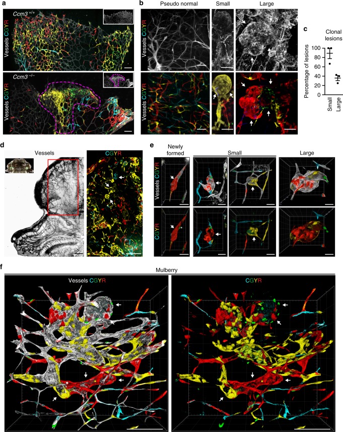
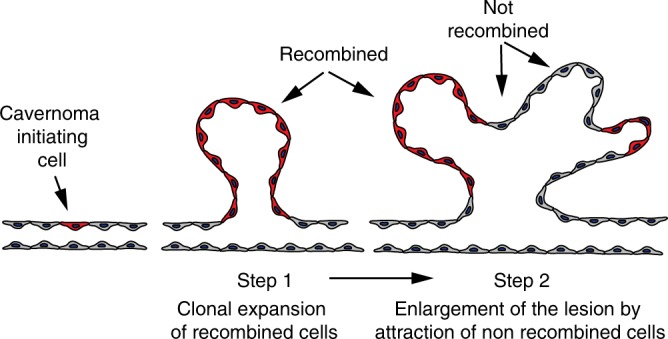
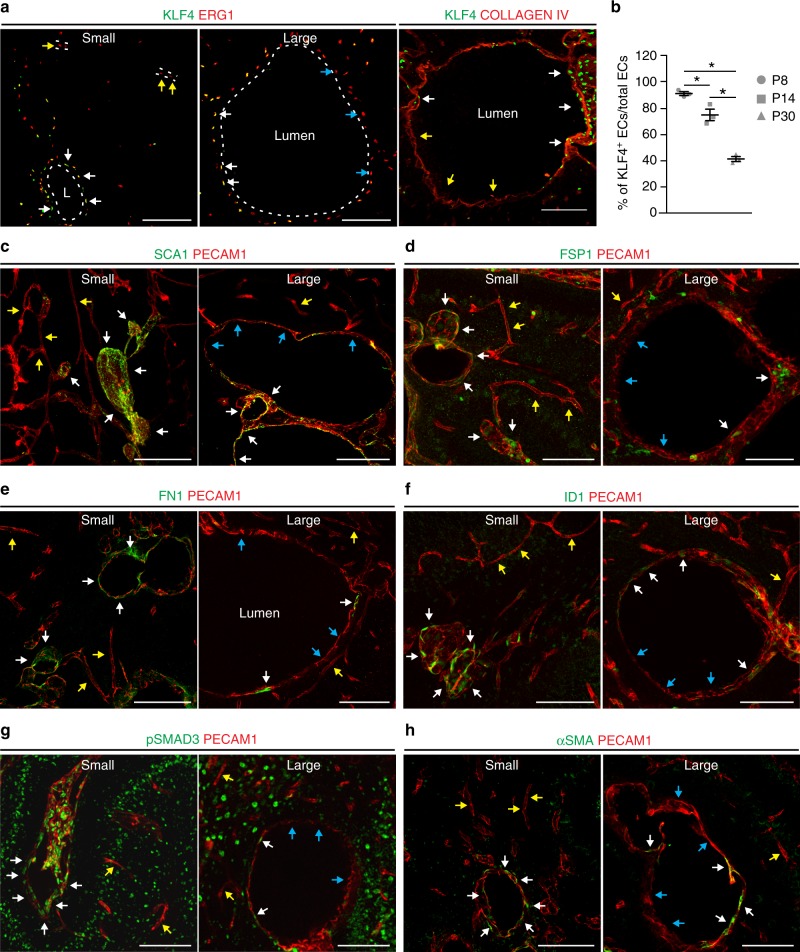
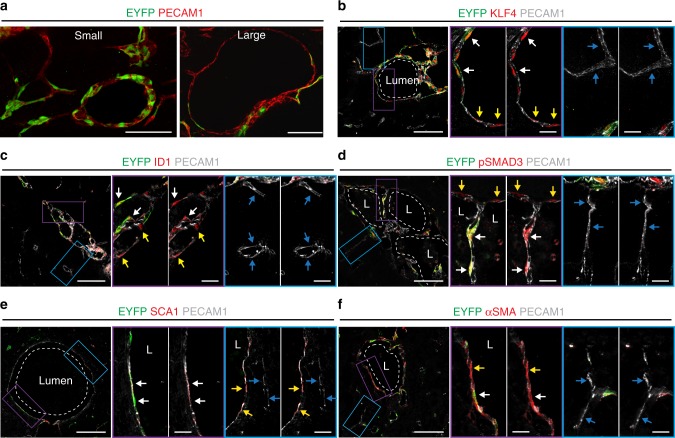
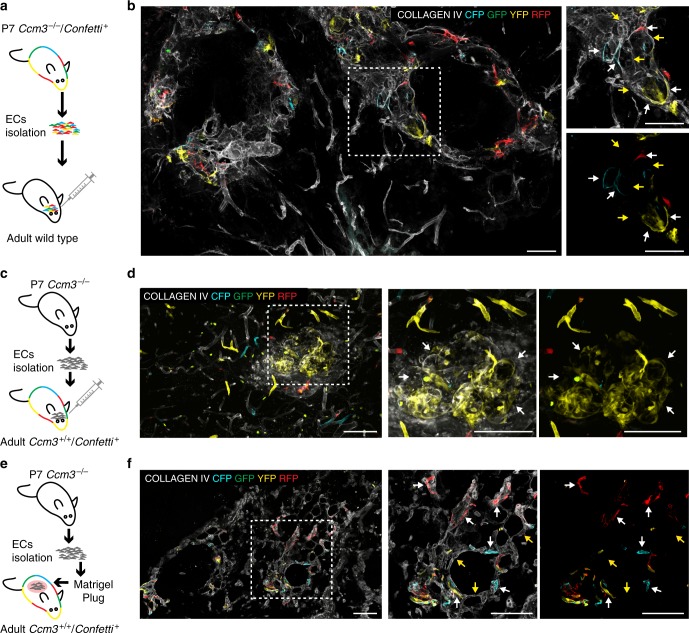
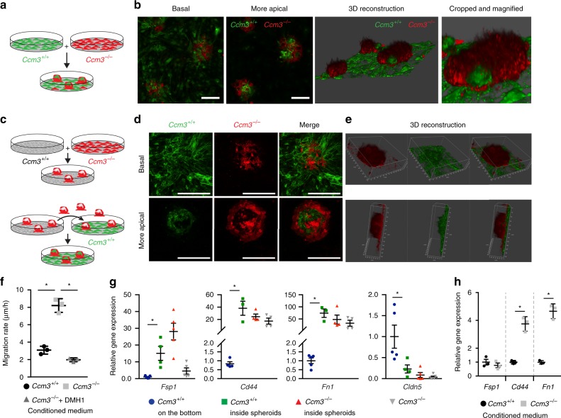
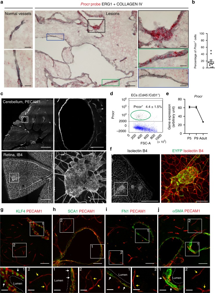
Cerebral cavernous malformation (CCM) is a neurovascular familial or sporadic disease that is characterised by capillary-venous cavernomas, and is due to loss-of-function mutations to any one of three CCM genes. Familial CCM follows a two-hit mechanism similar to that of tumour suppressor genes, while in sporadic cavernomas only a small fraction of endothelial cells shows mutated CCM genes. We reported that in mouse models and in human patients, endothelial cells lining the lesions have different features from the surrounding endothelium, as they express mesenchymal/stem-cell markers. Here we show that cavernomas originate from clonal expansion of few Ccm3-null endothelial cells that express mesenchymal/stem-cell markers. These cells then attract surrounding wild-type endothelial cells, inducing them to express mesenchymal/stem-cell markers and to contribute to cavernoma growth. These characteristics of Ccm3-null cells are reminiscent of the tumour-initiating cells that are responsible for tumour growth. Our data support the concept that CCM has benign tumour characteristics.

摘要

脑内海绵状血管畸形(CCM)是一种神经血管性家族性或散发性疾病,其特征为毛细血管-静脉海绵状畸形,是由于任何一个 CCM 基因的功能丧失性突变所致。家族性 CCM 遵循类似于肿瘤抑制基因的“双打击”机制,而在散发性海绵状血管畸形中,只有一小部分内皮细胞显示突变的 CCM 基因。我们曾报道,在小鼠模型和人类患者中,病变处的内皮细胞与周围内皮细胞具有不同的特征,因为它们表达间充质/干细胞标志物。在这里,我们显示海绵状血管畸形起源于少数表达间充质/干细胞标志物的 Ccm3 缺失内皮细胞的克隆扩张。这些细胞随后吸引周围的野生型内皮细胞,诱导它们表达间充质/干细胞标志物,并促进海绵状血管畸形的生长。Ccm3 缺失细胞的这些特征使人联想到负责肿瘤生长的肿瘤起始细胞。我们的数据支持 CCM 具有良性肿瘤特征的概念。

https://cdn.ncbi.nlm.nih.gov/pmc/blobs/8d0c/6591323/40aa0b8e88f8/41467_2019_10707_Fig9_HTML.jpg
https://cdn.ncbi.nlm.nih.gov/pmc/blobs/8d0c/6591323/03f9c98905dc/41467_2019_10707_Fig1_HTML.jpg
https://cdn.ncbi.nlm.nih.gov/pmc/blobs/8d0c/6591323/260130db9165/41467_2019_10707_Fig2_HTML.jpg
https://cdn.ncbi.nlm.nih.gov/pmc/blobs/8d0c/6591323/0af36b6a562c/41467_2019_10707_Fig3_HTML.jpg
https://cdn.ncbi.nlm.nih.gov/pmc/blobs/8d0c/6591323/f389a59d6e59/41467_2019_10707_Fig4_HTML.jpg
https://cdn.ncbi.nlm.nih.gov/pmc/blobs/8d0c/6591323/d0fa8fd01c7a/41467_2019_10707_Fig5_HTML.jpg
https://cdn.ncbi.nlm.nih.gov/pmc/blobs/8d0c/6591323/178c4e490eab/41467_2019_10707_Fig6_HTML.jpg
https://cdn.ncbi.nlm.nih.gov/pmc/blobs/8d0c/6591323/6373169b5435/41467_2019_10707_Fig7_HTML.jpg
https://cdn.ncbi.nlm.nih.gov/pmc/blobs/8d0c/6591323/a971a3cf7d52/41467_2019_10707_Fig8_HTML.jpg
https://cdn.ncbi.nlm.nih.gov/pmc/blobs/8d0c/6591323/40aa0b8e88f8/41467_2019_10707_Fig9_HTML.jpg

相似文献

1
Endothelial cell clonal expansion in the development of cerebral cavernous malformations.脑动静脉畸形发生中内皮细胞的克隆性扩张。
Nat Commun. 2019 Jun 24;10(1):2761. doi: 10.1038/s41467-019-10707-x.
2
Mural Cell-Specific Deletion of Cerebral Cavernous Malformation 3 in the Brain Induces Cerebral Cavernous Malformations.脑内血管性腔隙畸形 3 的壁细胞特异性缺失可诱导脑内血管性腔隙畸形。
Arterioscler Thromb Vasc Biol. 2020 Sep;40(9):2171-2186. doi: 10.1161/ATVBAHA.120.314586. Epub 2020 Jul 9.
3
Exceptional aggressiveness of cerebral cavernous malformation disease associated with PDCD10 mutations.与PDCD10突变相关的脑海绵状血管畸形疾病的异常侵袭性。
Genet Med. 2015 Mar;17(3):188-196. doi: 10.1038/gim.2014.97. Epub 2014 Aug 14.
4
Differential angiogenesis function of CCM2 and CCM3 in cerebral cavernous malformations.CCM2 和 CCM3 在脑动静脉畸形中的差异血管生成功能。
Neurosurg Focus. 2010 Sep;29(3):E1. doi: 10.3171/2010.5.FOCUS1090.
5
Endothelial Cells Lining Sporadic Cerebral Cavernous Malformation Cavernomas Undergo Endothelial-to-Mesenchymal Transition.散发性脑海绵状血管畸形(脑海绵状瘤)内的内皮细胞经历内皮-间充质转化。
Stroke. 2016 Mar;47(3):886-90. doi: 10.1161/STROKEAHA.115.011867. Epub 2016 Feb 2.
6
Apoptotic functions of PDCD10/CCM3, the gene mutated in cerebral cavernous malformation 3.PDCD10/CCM3的凋亡功能,该基因在脑海绵状血管畸形3中发生突变
Stroke. 2009 Apr;40(4):1474-81. doi: 10.1161/STROKEAHA.108.527135. Epub 2009 Feb 26.
7
Lesions from patients with sporadic cerebral cavernous malformations harbor somatic mutations in the CCM genes: evidence for a common biochemical pathway for CCM pathogenesis.散发性脑海绵状血管畸形患者的病灶中CCM基因存在体细胞突变:CCM发病机制的共同生化途径的证据
Hum Mol Genet. 2014 Aug 15;23(16):4357-70. doi: 10.1093/hmg/ddu153. Epub 2014 Apr 3.
8
Genomic causes of multiple cerebral cavernous malformations in a Japanese population.日本人多发性脑内海绵状血管畸形的基因组病因。
J Clin Neurosci. 2013 May;20(5):667-9. doi: 10.1016/j.jocn.2012.05.041. Epub 2013 Feb 26.
9
CCM3 and cerebral cavernous malformation disease.CCM3 与脑静脉血管畸形病。
Stroke Vasc Neurol. 2019 Mar 2;4(2):67-70. doi: 10.1136/svn-2018-000195. eCollection 2019 Jul.
10
Notch Signaling in Familial Cerebral Cavernous Malformations and Immunohistochemical Detection of Cleaved Notch1 Intracellular Domain.家族性脑海绵状畸形中的Notch信号通路及切割的Notch1细胞内结构域的免疫组织化学检测
Methods Mol Biol. 2020;2152:427-435. doi: 10.1007/978-1-0716-0640-7_31.

引用本文的文献

1
Semaphorin 3A and 3F overexpression in TIE2 hyperactive endothelial cells contribute to the pathological lumen expansion in venous malformation.在TIE2过度活跃的内皮细胞中,信号素3A和3F的过表达促成了静脉畸形中的病理性管腔扩张。
bioRxiv. 2025 Jul 24:2025.07.18.665640. doi: 10.1101/2025.07.18.665640.
2
The Role of Somatic Mutation in Hereditary Hemorrhagic Telangiectasia Pathogenesis.体细胞突变在遗传性出血性毛细血管扩张症发病机制中的作用。
J Clin Med. 2025 Jun 24;14(13):4479. doi: 10.3390/jcm14134479.
3
Single-cell transcriptome profiling reveals dynamic cell populations and immune infiltration in cerebral cavernous malformation.

本文引用的文献

1
Fine-Tuning of Sox17 and Canonical Wnt Coordinates the Permeability Properties of the Blood-Brain Barrier.Sox17 和经典 Wnt 的精细调控协调血脑屏障的通透性特性。
Circ Res. 2019 Feb 15;124(4):511-525. doi: 10.1161/CIRCRESAHA.118.313316.
2
Endothelial cell transitions.内皮细胞转变
Science. 2018 Nov 16;362(6416):746-747. doi: 10.1126/science.aas9432.
3
Cerebral Cavernous Malformations Develop Through Clonal Expansion of Mutant Endothelial Cells.脑内海绵状血管畸形通过突变内皮细胞的克隆扩张发展。
单细胞转录组分析揭示了脑海绵状血管畸形中的动态细胞群和免疫浸润。
Front Immunol. 2025 May 30;16:1592343. doi: 10.3389/fimmu.2025.1592343. eCollection 2025.
4
High-throughput differentiation of human blood vessel organoids reveals overlapping and distinct functions of the cerebral cavernous malformation proteins.人血管类器官的高通量分化揭示了脑海绵状血管畸形蛋白的重叠和独特功能。
Angiogenesis. 2025 Jun 6;28(3):32. doi: 10.1007/s10456-025-09985-5.
5
Focused ultrasound-microbubble treatment arrests the growth and formation of cerebral cavernous malformations.聚焦超声-微泡治疗可抑制脑海绵状血管畸形的生长和形成。
Nat Biomed Eng. 2025 May 13. doi: 10.1038/s41551-025-01390-z.
6
Pharmacological blocking of neutrophil extracellular traps attenuates immunothrombosis and neuroinflammation in cerebral cavernous malformation.中性粒细胞胞外诱捕网的药理学阻断可减轻海绵状血管瘤中的免疫血栓形成和神经炎症。
Nat Cardiovasc Res. 2024 Dec;3(12):1549-1567. doi: 10.1038/s44161-024-00577-y. Epub 2024 Dec 4.
7
Advances in regulating endothelial-mesenchymal transformation through exosomes.外泌体在调控血管内皮-间质转化中的研究进展。
Stem Cell Res Ther. 2024 Oct 31;15(1):391. doi: 10.1186/s13287-024-04010-w.
8
Force-mediated recruitment and reprogramming of healthy endothelial cells drive vascular lesion growth.力介导的健康内皮细胞募集和重编程驱动血管病变生长。
Nat Commun. 2024 Oct 6;15(1):8660. doi: 10.1038/s41467-024-52866-6.
9
PDCD10 Is a Key Player in TMZ-Resistance and Tumor Cell Regrowth: Insights into Its Underlying Mechanism in Glioblastoma Cells.程序性死亡配体 10 是 TMZ 耐药和肿瘤细胞再生的关键因素:其在胶质母细胞瘤细胞中的潜在机制的见解。
Cells. 2024 Aug 28;13(17):1442. doi: 10.3390/cells13171442.
10
Pathophysiology in Brain Arteriovenous Malformations: Focus on Endothelial Dysfunctions and Endothelial-to-Mesenchymal Transition.脑动静脉畸形的病理生理学:聚焦于内皮功能障碍和内皮-间充质转化
Biomedicines. 2024 Aug 7;12(8):1795. doi: 10.3390/biomedicines12081795.
Circ Res. 2018 Oct 26;123(10):1143-1151. doi: 10.1161/CIRCRESAHA.118.313970.
4
CD157 Marks Tissue-Resident Endothelial Stem Cells with Homeostatic and Regenerative Properties.CD157 标记具有稳态和再生特性的组织驻留内皮干细胞。
Cell Stem Cell. 2018 Mar 1;22(3):384-397.e6. doi: 10.1016/j.stem.2018.01.010. Epub 2018 Feb 8.
5
Clonal Expansion of Endothelial Cells Contributes to Ischemia-Induced Neovascularization.内皮细胞的克隆扩增有助于缺血诱导的血管新生。
Circ Res. 2018 Mar 2;122(5):670-677. doi: 10.1161/CIRCRESAHA.117.312310. Epub 2018 Jan 22.
6
EMT in cancer.肿瘤中的 EMT。
Nat Rev Cancer. 2018 Feb;18(2):128-134. doi: 10.1038/nrc.2017.118. Epub 2018 Jan 12.
7
Deregulated TGF-β/BMP Signaling in Vascular Malformations.血管畸形中 TGF-β/BMP 信号的失调。
Circ Res. 2017 Sep 29;121(8):981-999. doi: 10.1161/CIRCRESAHA.117.309930.
8
Role of bone morphogenetic proteins in sprouting angiogenesis: differential BMP receptor-dependent signaling pathways balance stalk tip cell competence.骨形态发生蛋白在发芽血管生成中的作用:不同的骨形态发生蛋白受体依赖性信号通路平衡茎尖细胞能力。
FASEB J. 2017 Nov;31(11):4720-4733. doi: 10.1096/fj.201700193RR. Epub 2017 Jul 21.
9
EMT, CSCs, and drug resistance: the mechanistic link and clinical implications.上皮-间质转化、癌症干细胞与耐药性:机制联系及临床意义
Nat Rev Clin Oncol. 2017 Oct;14(10):611-629. doi: 10.1038/nrclinonc.2017.44. Epub 2017 Apr 11.
10
Alk2/ACVR1 and Alk3/BMPR1A Provide Essential Function for Bone Morphogenetic Protein-Induced Retinal Angiogenesis.Alk2/ACVR1和Alk3/BMPR1A对骨形态发生蛋白诱导的视网膜血管生成具有重要作用。
Arterioscler Thromb Vasc Biol. 2017 Apr;37(4):657-663. doi: 10.1161/ATVBAHA.116.308422. Epub 2017 Feb 23.