Institute for Biomedical Engineering and Institute of Pharmacology and Toxicology, University of Zurich and ETH Zurich, Zurich, Switzerland.
Institute for Biological and Medical Imaging, Technical University of Munich and Helmholtz Center Munich, Neuherberg, Germany; iThera Medical GmbH, Munich, Germany.
Neoplasia. 2020 Dec;22(12):770-777. doi: 10.1016/j.neo.2020.10.008. Epub 2020 Nov 1.
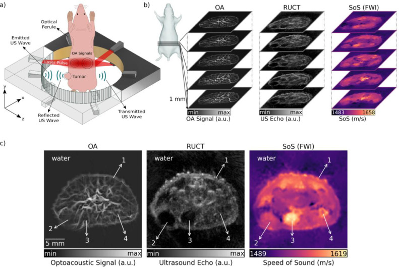
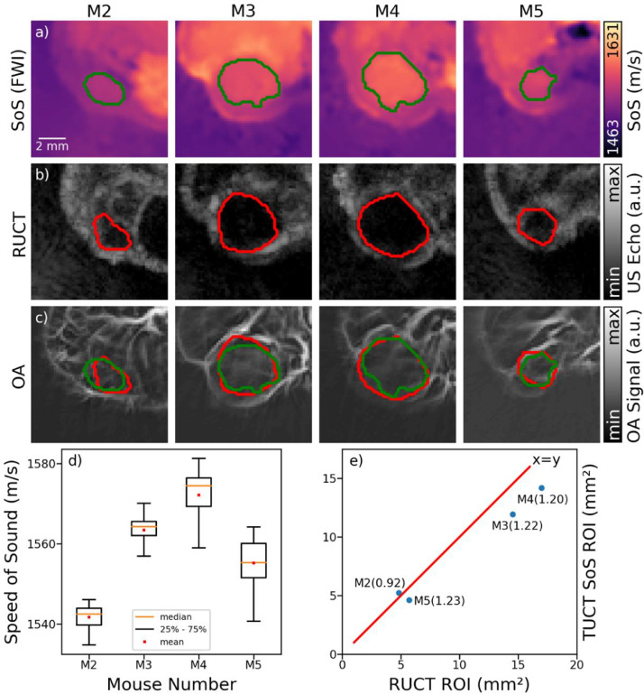
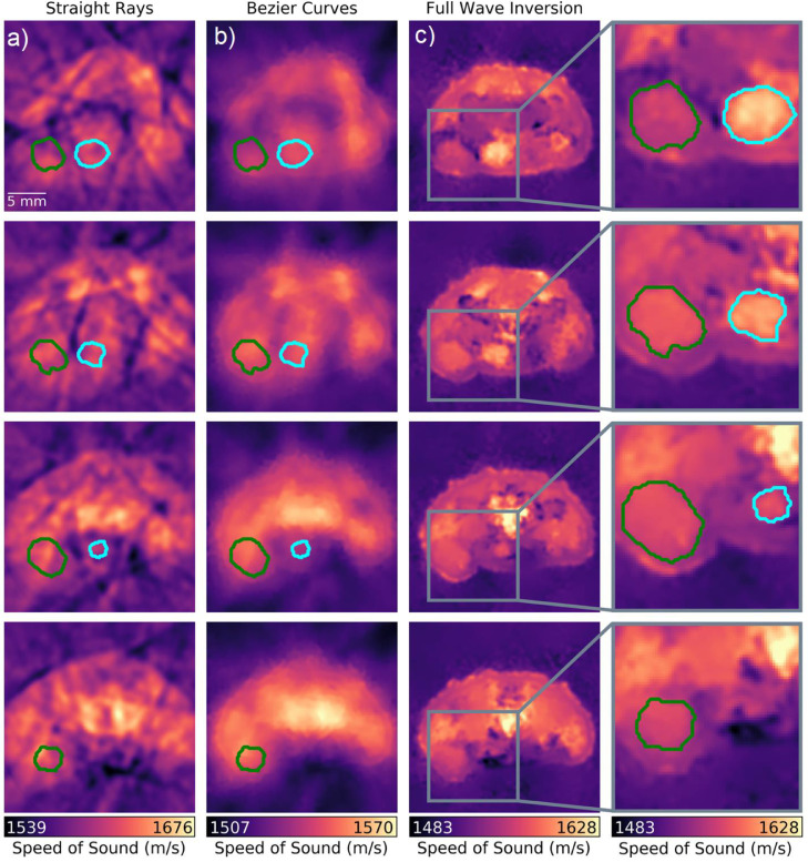
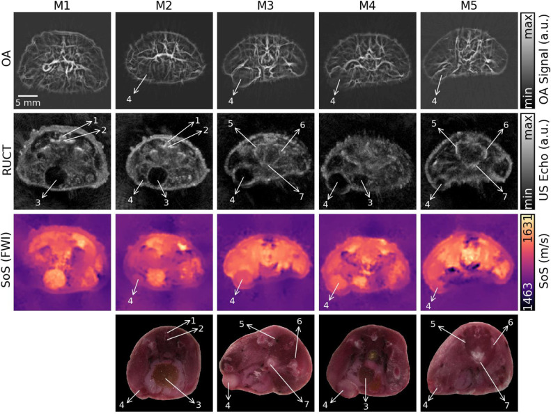
Development of imaging methods capable of furnishing tumor-specific morphological, functional, and molecular information is paramount for early diagnosis, staging, and treatment of breast cancer. Ultrasound (US) and optoacoustic (OA) imaging methods exhibit excellent traits for tumor imaging in terms of fast imaging speed, ease of use, excellent contrast, and lack of ionizing radiation. Here, we demonstrate simultaneous tomographic whole body imaging of optical absorption, US reflectivity, and speed of sound (SoS) in living mice. In vivo studies of 4T1 breast cancer xenografts models revealed synergistic and complementary value of the hybrid imaging approach for characterizing mammary tumors. While neovasculature surrounding the tumor areas were observed based on the vascular anatomy contrast provided by the OA data, the tumor boundaries could be discerned by segmenting hypoechoic structures in pulse-echo US images. Tumor delineation was further facilitated by enhancing the contrast and spatial resolution of the SoS maps with a full-wave inversion method. The malignant lesions could thus be distinguished from other hypoechoic regions based on the average SoS values. The reported findings corroborate the strong potential of the hybrid imaging approach for advancing cancer research in small animal models and fostering development of new clinical diagnostic approaches.
开发能够提供肿瘤特异性形态、功能和分子信息的成像方法对于乳腺癌的早期诊断、分期和治疗至关重要。超声(US)和光声(OA)成像方法在肿瘤成像方面具有出色的特性,包括快速成像速度、易于使用、出色的对比度和无电离辐射。在这里,我们展示了在活体小鼠中同时进行光学吸收、US 反射率和声速(SoS)的层析全身体成像。在 4T1 乳腺癌异种移植模型的体内研究中,该混合成像方法在表征乳腺肿瘤方面具有协同和互补的价值。虽然可以根据 OA 数据提供的血管解剖对比观察到肿瘤区域周围的新生血管,但可以通过对脉冲回波 US 图像中的低回声结构进行分割来辨别肿瘤边界。通过使用全波反演方法增强 SoS 图谱的对比度和空间分辨率,进一步促进了肿瘤的描绘。因此,可以基于平均 SoS 值将恶性病变与其他低回声区域区分开来。所报道的发现证实了混合成像方法在推进小动物模型中的癌症研究和促进新的临床诊断方法发展方面的强大潜力。