Juříková Tereza, Luptáková Dominika, Kofroňová Olga, Škríba Anton, Novák Jiří, Marešová Helena, Palyzová Andrea, Petřík Miloš, Havlíček Vladimír, Benada Oldřich
Institute of Microbiology of the Czech Academy of Sciences, Vídeňská 1083, 142 20 Prague, Czech Republic.
Department of Genetics and Microbiology, Faculty of Science, Charles University, Viničná 5, 128 44 Prague, Czech Republic.
J Fungi (Basel). 2020 Oct 30;6(4):257. doi: 10.3390/jof6040257.
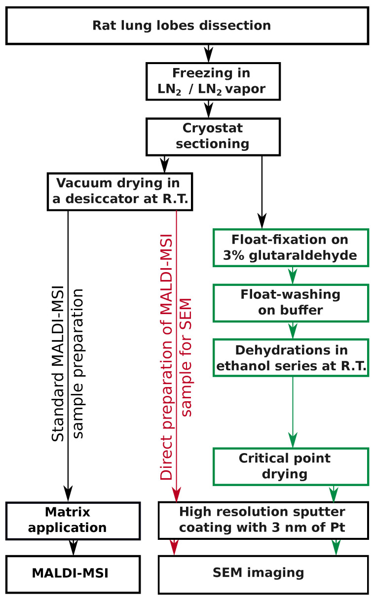
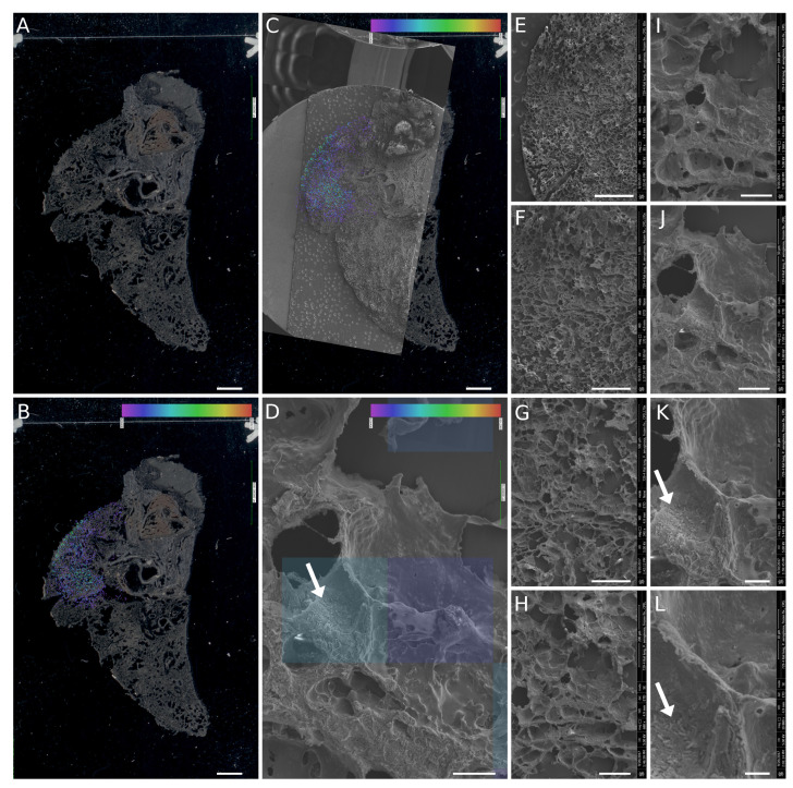
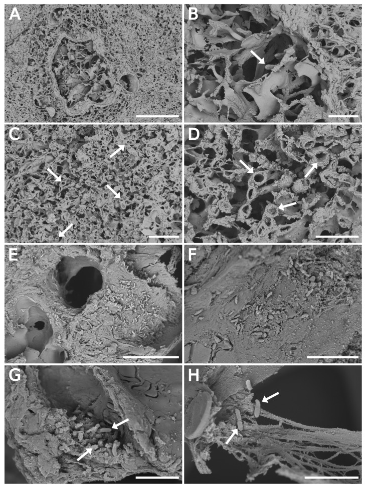
A procedure for processing frozen rat lung tissue sections for scanning electron microscopy (SEM) from deeply frozen samples initially collected and stored for matrix-assisted laser desorption/ionization mass spectrometry imaging (MALDI-MSI) was developed. The procedure employed slow thawing of the frozen sections while floating on the surface and melting in a fixative solution. After the float-washing step, the sections were dehydrated in a graded ethanol series and dried in a critical point dryer. The SEM generated images with well-preserved structures, allowing for monitoring of bacterial cells and fungal hyphae in the infected tissue. Importantly, the consecutive nonfixed frozen sections were fully compatible with MALDI-MSI, providing molecular biomarker maps of . The protocol enables bimodal image fusion in the in-house software CycloBranch, as demonstrated by SEM and MALDI-MSI.
开发了一种用于处理冷冻大鼠肺组织切片的程序,该程序用于对最初收集并储存用于基质辅助激光解吸/电离质谱成像(MALDI-MSI)的深度冷冻样本进行扫描电子显微镜(SEM)观察。该程序采用将冷冻切片在固定液表面缓慢解冻并融化。在漂浮冲洗步骤之后,切片在梯度乙醇系列中脱水,并在临界点干燥器中干燥。扫描电子显微镜生成的图像结构保存完好,能够监测感染组织中的细菌细胞和真菌菌丝。重要的是,连续的未固定冷冻切片与基质辅助激光解吸/电离质谱成像完全兼容,可提供……的分子生物标志物图谱。如扫描电子显微镜和基质辅助激光解吸/电离质谱成像所示,该方案能够在内部软件CycloBranch中实现双峰图像融合。