Reategui Cesar
Missouri Delta Medical Center, General Surgery Department, 1008 N Main St, Sikeston, MO 63801, United States of America.
Trauma Case Rep. 2020 Oct 8;30:100362. doi: 10.1016/j.tcr.2020.100362. eCollection 2020 Dec.
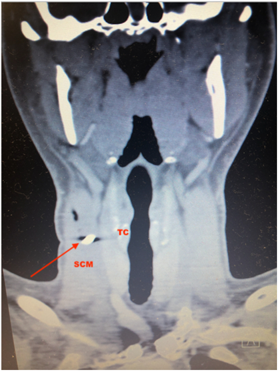
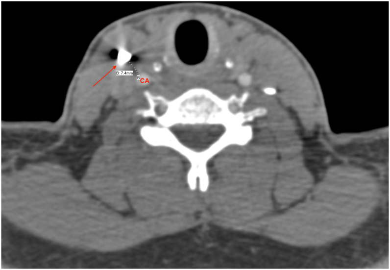
Penetrating neck wounds can be fatal and require prompt attention. The trauma literature is flooded with management protocols for penetrating wounds to the neck; however, in the absence of hard signs the definitive management of lodged foreign bodies beyond the platysma is less clear. This report describes a work-related injury of a Caucasian 33-year-old male who arrived in the Emergency Department (ER) with a 1 cm metallic foreign body (FB) lodged in zone II of the neck, 7 mm antero-lateral to the right internal carotid artery. The technical aspects of its retrieval are discussed as well as a literature review of the current management of embedded FBs in the neck. The patient was taken to the operating room and the FB was removed via a 3 cm incision. Fluoroscopy was used for exact localization of and to allow a precise skin incision overlying the FB. The FB was retrieved uneventfully; a fiberoptic esophagoscopy and bronchoscopy showed normal findings. The patient was discharged home the next day. At 15 months follow-up he is doing well without sequela. The use of fluoroscopy is strongly encouraged for FB removal in asymptomatic patients. The management of lodged foreign bodies in the neck should be part of future guidelines.
颈部穿透伤可能致命,需要及时处理。创伤文献中充斥着颈部穿透伤的处理方案;然而,在没有明确体征的情况下,对于位于颈阔肌以下的异物的最终处理尚不清楚。本报告描述了一名33岁白种男性的工伤,他因一枚1厘米的金属异物嵌入颈部II区、右颈内动脉前外侧7毫米处而被送往急诊科。文中讨论了取出异物的技术要点,并对目前颈部嵌入异物的处理进行了文献综述。患者被送往手术室,通过一个3厘米的切口取出了异物。使用荧光透视法进行精确定位,并在异物上方进行精确的皮肤切口。异物顺利取出;纤维食管镜和支气管镜检查结果正常。患者第二天出院。在15个月的随访中,他情况良好,没有后遗症。强烈建议对无症状患者使用荧光透视法取出异物。颈部异物的处理应成为未来指南的一部分。