Ramseyer Abigail M, Dajani Nafisa
Department of Obstetrics & Gynecology, Division of Maternal Fetal Medicine, University of Arkansas for Medical Sciences, Little Rock, AR 72205, United States of America.
Case Rep Womens Health. 2020 Oct 17;28:e00266. doi: 10.1016/j.crwh.2020.e00266. eCollection 2020 Oct.
Split notochord syndrome is a rare disorder characterized by fistula formation between the gastrointestinal tract and skin on the dorsum. Prenatal diagnosis is difficult and most cases are diagnosed postnatally.
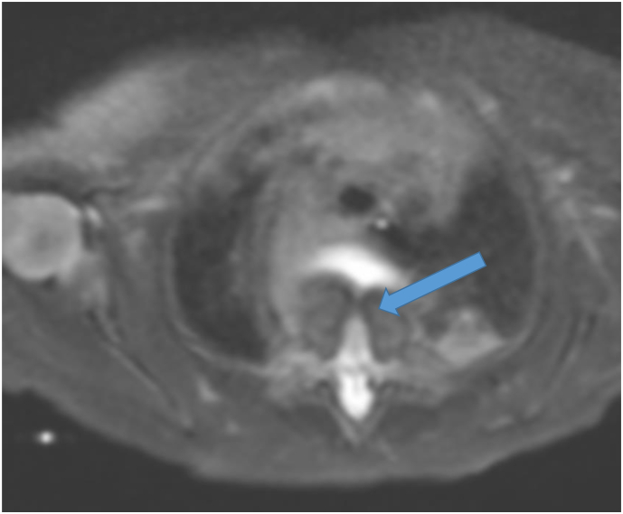
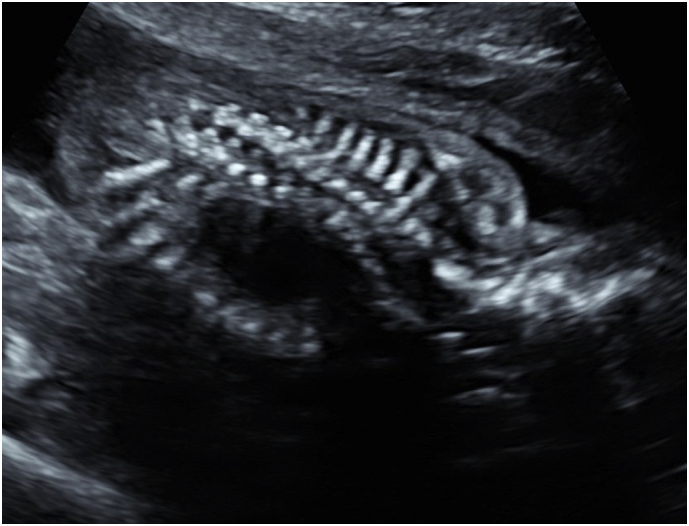
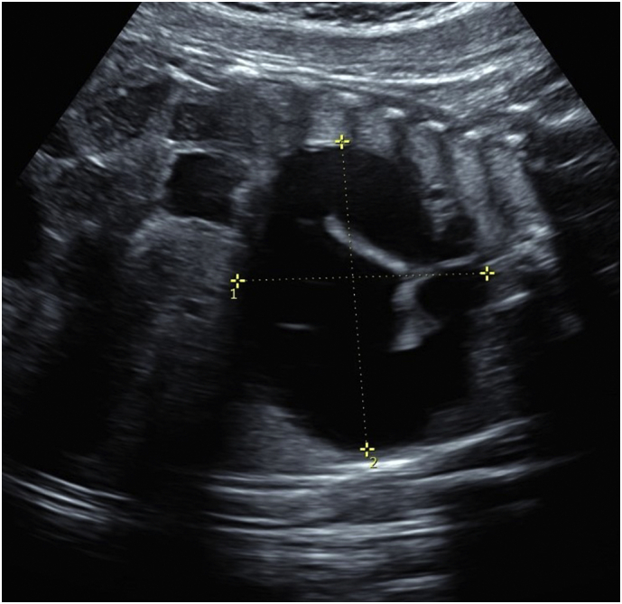
A 29-year-old woman, gravida 3 para 2, was referred for fetal cystic chest mass on prenatal ultrasound for congenital pulmonary airway malformation (CPAM). Fetal magnetic resonance imaging (MRI) suggested foregut duplication, and this was confirmed on postnatal thoracotomy with mass excision. A spine dysraphism was suspected on prenatal ultrasound, but was not confirmed on fetal MRI at the time of the study. Neonatal MRI noted vertebral abnormalities, confirming split notochord syndrome. Retrospective examination of the fetal MRI images detected a dysraphism and confirmed the prenatal ultrasound findings.
At 17 months of life, the child had mild symptoms of neurogenic bowel, but was meeting all milestones without neurodevelopmental delays. We present a mild form of split notochord syndrome.
Split notochord syndrome is difficult to diagnose prenatally and should be considered when a fetal cystic chest mass is found on ultrasound. Detailed vertebrae assessment may improve detection.
背侧脊索分裂综合征是一种罕见疾病,其特征为胃肠道与背部皮肤之间形成瘘管。产前诊断困难,大多数病例在出生后才得以确诊。
一名29岁女性,孕3产2,因产前超声检查发现胎儿胸部囊性肿块,诊断为先天性肺气道畸形(CPAM)而前来就诊。胎儿磁共振成像(MRI)提示前肠重复畸形,产后开胸切除肿块时得以证实。产前超声怀疑脊柱裂,但在研究时胎儿MRI未得到证实。新生儿MRI发现椎体异常,确诊为背侧脊索分裂综合征。对胎儿MRI图像进行回顾性检查发现脊柱裂,证实了产前超声检查结果。
患儿17个月大时,有轻度神经源性肠道症状,但各项发育指标均正常,无神经发育迟缓。我们报告了一例轻度背侧脊索分裂综合征病例。
背侧脊索分裂综合征产前诊断困难,超声检查发现胎儿胸部囊性肿块时应考虑该病。详细的椎体评估可能会提高检出率。