• 文献检索
  • 文档翻译
  • 深度研究
  • 学术资讯
  • Suppr Zotero 插件Zotero 插件
  • 邀请有礼
  • 套餐&价格
  • 历史记录
应用&插件
Suppr Zotero 插件Zotero 插件浏览器插件Mac 客户端Windows 客户端微信小程序
定价
高级版会员购买积分包购买API积分包
服务
文献检索文档翻译深度研究API 文档MCP 服务
关于我们
关于 Suppr公司介绍联系我们用户协议隐私条款
关注我们

Suppr 超能文献

核心技术专利:CN118964589B侵权必究
粤ICP备2023148730 号-1Suppr @ 2026

文献检索

告别复杂PubMed语法,用中文像聊天一样搜索,搜遍4000万医学文献。AI智能推荐,让科研检索更轻松。

立即免费搜索

文件翻译

保留排版,准确专业,支持PDF/Word/PPT等文件格式,支持 12+语言互译。

免费翻译文档

深度研究

AI帮你快速写综述,25分钟生成高质量综述,智能提取关键信息,辅助科研写作。

立即免费体验

皮肤镜下彩虹模式:恶性肿瘤的有力线索还是只是一场光影秀?

Dermoscopic rainbow pattern: A strong clue to malignancy or just a light show?

作者信息

Elmas Omer Faruk, Mayisoglu Herman, Celik Murat, Kilitci Asuman, Akdeniz Necmettin

机构信息

Department of Dermatology and Veneorology, Ahi Evran University Faculty of Medicine, Kirsehir, Turkey.

Department of Dermatology and Veneorology, Medicana Hospital, Istanbul, Turkey.

出版信息

North Clin Istanb. 2020 Aug 31;7(5):494-498. doi: 10.14744/nci.2020.32656. eCollection 2020.

DOI:10.14744/nci.2020.32656
PMID:33163886
原文链接:https://pmc.ncbi.nlm.nih.gov/articles/PMC7603844/
Abstract

OBJECTIVE

Rainbow pattern is a dermoscopic finding composed of multiple colors simulating a rainbow. It is known as a characteristic feature of Kaposi's sarcoma. Here, we reported different non-Kaposi's sarcoma conditions with a rainbow pattern aiming to discuss the diagnostic significance of the finding.

METHODS

In this multicenter study, dermoscopic images of the non-Kaposi's sarcoma lesions having a histopathological diagnosis were reviewed for the presence of a rainbow pattern. Dermoscopic examination was performed by a polarized handheld dermoscope with x10 magnification.

RESULTS

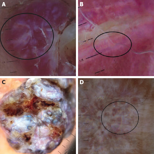
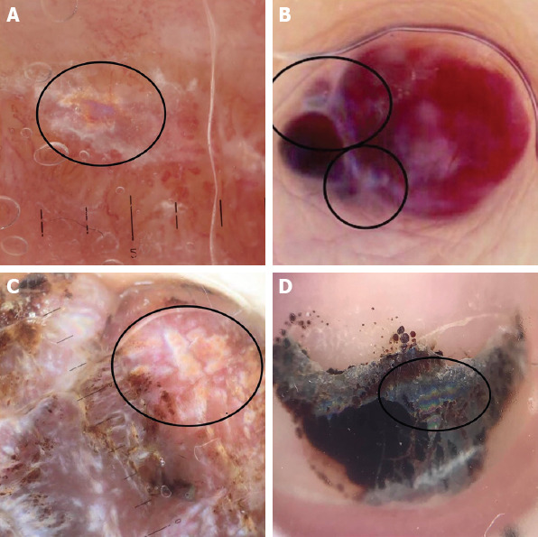
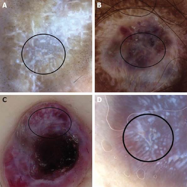
A total of 840 lesions were reviewed and 21 (2%) non-Kaposi sarcoma lesions having dermoscopic rainbow pattern were detected. These lesions were as follows; pyogenic granuloma (n=4, 19%), hypertrophic scar (n=4, 19%), basal cell carcinoma (n=2, 10%), dermatofibroma (n=2, 10%), angiokeratoma (n=2, 10%), blue nevus (n=1, 5%), granuloma annulare (n=1, 5%), strawberry angioma (n=1, 5%), epidermal cyst (n=1, 5%), malignant melanoma (n=1, 5%), dissecting cellulitis (n=1, 5%) and subungual hematoma (n=1, 5%). The most common localization was limb (n=14, 67%) followed by face (n=3, 14%).

CONCLUSION

We suggest that the rainbow pattern is a complex and quite unspecific optic phenomenon which can be seen both in vascular and non-vascular lesions. Its diagnostic significance should be considered in the context of the other structural dermoscopic finding. To the best of our knowledge, to our knowledge, this is the most comprehensive study focusing on rainbow pattern in non-Kaposi's sarcoma lesions. Here, we also reported rainbow pattern in dissecting cellulitis, granuloma annulare and subungual hematoma which has not been shown to have rainbow pattern previously.

摘要

目的

彩虹样模式是一种皮肤镜表现,由多种颜色组成,形似彩虹。它是卡波西肉瘤的一个特征性表现。在此,我们报告了具有彩虹样模式的不同非卡波西肉瘤情况,旨在探讨该表现的诊断意义。

方法

在这项多中心研究中,对具有组织病理学诊断的非卡波西肉瘤病变的皮肤镜图像进行回顾,以检查是否存在彩虹样模式。使用带10倍放大倍数的偏振手持式皮肤镜进行皮肤镜检查。

结果

共回顾了840个病变,检测到21个(2%)具有皮肤镜彩虹样模式的非卡波西肉瘤病变。这些病变如下:化脓性肉芽肿(n = 4,19%)、肥厚性瘢痕(n = 4,19%)、基底细胞癌(n = 2,10%)、皮肤纤维瘤(n = 2,10%)、血管角皮瘤(n = 2,10%)、蓝痣(n = 1,5%)、环状肉芽肿(n = 1,5%)、草莓状血管瘤(n = 1,5%)、表皮囊肿(n = 1,5%)、恶性黑色素瘤(n = 1,5%)、蜂窝织炎(n = 1,5%)和甲下血肿(n = 1,5%)。最常见的部位是四肢(n = 14,67%),其次是面部(n = 3,14%)。

结论

我们认为彩虹样模式是一种复杂且相当非特异性的光学现象,在血管性和非血管性病变中均可出现。其诊断意义应结合其他皮肤镜结构表现来考虑。据我们所知,这是关于非卡波西肉瘤病变中彩虹样模式的最全面研究。在此,我们还报告了蜂窝织炎、环状肉芽肿和甲下血肿中的彩虹样模式,此前尚未显示这些病变有彩虹样模式。

https://cdn.ncbi.nlm.nih.gov/pmc/blobs/7926/7603844/ebc1321eee79/NCI-7-494-g003.jpg
https://cdn.ncbi.nlm.nih.gov/pmc/blobs/7926/7603844/2c8519976b6a/NCI-7-494-g001.jpg
https://cdn.ncbi.nlm.nih.gov/pmc/blobs/7926/7603844/ee7fd3800e1d/NCI-7-494-g002.jpg
https://cdn.ncbi.nlm.nih.gov/pmc/blobs/7926/7603844/ebc1321eee79/NCI-7-494-g003.jpg
https://cdn.ncbi.nlm.nih.gov/pmc/blobs/7926/7603844/2c8519976b6a/NCI-7-494-g001.jpg
https://cdn.ncbi.nlm.nih.gov/pmc/blobs/7926/7603844/ee7fd3800e1d/NCI-7-494-g002.jpg
https://cdn.ncbi.nlm.nih.gov/pmc/blobs/7926/7603844/ebc1321eee79/NCI-7-494-g003.jpg

相似文献

1
Dermoscopic rainbow pattern: A strong clue to malignancy or just a light show?皮肤镜下彩虹模式:恶性肿瘤的有力线索还是只是一场光影秀?
North Clin Istanb. 2020 Aug 31;7(5):494-498. doi: 10.14744/nci.2020.32656. eCollection 2020.
2
Dermoscopy of Kaposi's sarcoma: areas exhibiting the multicoloured 'rainbow pattern'.卡波西肉瘤的皮肤镜检查:呈现多色“彩虹图案”的区域。
J Eur Acad Dermatol Venereol. 2009 Oct;23(10):1128-32. doi: 10.1111/j.1468-3083.2009.03239.x. Epub 2009 Apr 10.
3
Rainbow pattern in Kaposi's sarcoma under polarized dermoscopy: a dermoscopic pathological study.偏振皮肤镜下卡波西肉瘤的彩虹图案:一项皮肤镜病理研究
Br J Dermatol. 2009 Apr;160(4):801-9. doi: 10.1111/j.1365-2133.2008.08940.x. Epub 2008 Dec 1.
4
The Dermoscopic Rainbow Pattern - A Review of the Literature.皮肤镜下的彩虹模式——文献综述
Acta Dermatovenerol Croat. 2019 Jun;27(2):111-115.
5
Pyogenic granuloma and nodular Kaposi’s sarcoma: dermoscopic clues for the differential diagnosis.化脓性肉芽肿和结节性卡波西肉瘤:鉴别诊断的皮肤镜线索。
Turk J Med Sci. 2019 Oct 24;49(5):1471-1478. doi: 10.3906/sag-1902-60.
6
The rainbow pattern in dermoscopy: A zoom on nonkaposi sarcoma skin diseases.皮肤镜下的彩虹图案:非卡波西肉瘤性皮肤病特写。
Biomed J. 2018 Jun;41(3):209-210. doi: 10.1016/j.bj.2018.04.004. Epub 2018 Jul 11.
7
Dermoscopic rainbow pattern in Kaposi's sarcoma lesions: our experience.卡波西肉瘤皮损的皮肤镜彩虹模式:我们的经验
Arch Dermatol. 2012 Oct;148(10):1207-8. doi: 10.1001/archdermatol.2012.2204.
8
'Rainbow pattern': a dermoscopic sign of invasive melanoma.“彩虹图案”:一种侵袭性黑色素瘤的皮肤镜征象。
Clin Exp Dermatol. 2022 Mar;47(3):529-533. doi: 10.1111/ced.14950. Epub 2021 Nov 10.
9
Dermoscopic diagnosis of subungual hematoma: new observations.甲下血肿的皮肤镜诊断:新观察结果
Postepy Dermatol Alergol. 2020 Aug;37(4):490-494. doi: 10.5114/ada.2020.98235. Epub 2020 Sep 2.
10
Clinical, Dermoscopic, Ultrasonographic, and Histopathologic Correlations in Kaposi's Sarcoma Lesions and Their Differential Diagnoses: A Single-Center Prospective Study.卡波西肉瘤病变的临床、皮肤镜、超声及组织病理学相关性及其鉴别诊断:一项单中心前瞻性研究
J Clin Med. 2022 Dec 29;12(1):278. doi: 10.3390/jcm12010278.

引用本文的文献

1
Non-Invasive Diagnostic Imaging in Kaposi Sarcoma Evaluation.卡波西肉瘤评估中的非侵入性诊断成像
Diagnostics (Basel). 2025 Jun 30;15(13):1665. doi: 10.3390/diagnostics15131665.
2
"Chasing Rainbows" Beyond Kaposi Sarcoma's Dermoscopy: A Mini-Review.《追逐彩虹》:超越卡波西肉瘤的皮肤镜检查——一篇综述
Dermatopathology (Basel). 2024 Nov 25;11(4):333-341. doi: 10.3390/dermatopathology11040035.
3
Iridescent Changes Observed During Dynamic Cross-polarized Dermoscopy.动态交叉偏振皮肤镜检查中观察到的彩虹色变化

本文引用的文献

1
Pyogenic granuloma and nodular Kaposi’s sarcoma: dermoscopic clues for the differential diagnosis.化脓性肉芽肿和结节性卡波西肉瘤:鉴别诊断的皮肤镜线索。
Turk J Med Sci. 2019 Oct 24;49(5):1471-1478. doi: 10.3906/sag-1902-60.
2
The rainbow pattern in dermoscopy: A zoom on nonkaposi sarcoma skin diseases.皮肤镜下的彩虹图案:非卡波西肉瘤性皮肤病特写。
Biomed J. 2018 Jun;41(3):209-210. doi: 10.1016/j.bj.2018.04.004. Epub 2018 Jul 11.
3
Basal cell carcinoma under the rainbow.彩虹下的基底细胞癌。
Dermatol Pract Concept. 2022 Jul 1;12(3):e2022119. doi: 10.5826/dpc.1203a119. eCollection 2022 Jul.
4
..
JAAD Case Rep. 2022 Jan 21;21:144-147. doi: 10.1016/j.jdcr.2022.01.001. eCollection 2022 Mar.
G Ital Dermatol Venereol. 2020 Apr;155(2):244-245. doi: 10.23736/S0392-0488.18.05968-0. Epub 2018 Apr 19.
4
Dermoscopic rainbow pattern in blue nevus.蓝痣的皮肤镜下彩虹模式。
Dermatol Pract Concept. 2017 Jul 31;7(3):60-62. doi: 10.5826/dpc.0703a13. eCollection 2017 Jul.
5
Image Gallery: Merkel cell carcinoma under the rainbow.图片库:彩虹下的默克尔细胞癌。
Br J Dermatol. 2017 Oct;177(4):e166. doi: 10.1111/bjd.15815.
6
Acral pseudolymphomatous angiokeratoma of children with rainbow pattern: A mimicker of Kaposi sarcoma.具有彩虹样图案的儿童肢端假淋巴瘤性血管角皮瘤:卡波西肉瘤的模仿者。
J Am Acad Dermatol. 2017 Feb;76(2S1):S25-S27. doi: 10.1016/j.jaad.2016.04.065.
7
Dermoscopic rainbow pattern in Kaposi's sarcoma lesions: our experience.卡波西肉瘤皮损的皮肤镜彩虹模式:我们的经验
Arch Dermatol. 2012 Oct;148(10):1207-8. doi: 10.1001/archdermatol.2012.2204.
8
Dermoscopy of Kaposi's sarcoma: areas exhibiting the multicoloured 'rainbow pattern'.卡波西肉瘤的皮肤镜检查:呈现多色“彩虹图案”的区域。
J Eur Acad Dermatol Venereol. 2009 Oct;23(10):1128-32. doi: 10.1111/j.1468-3083.2009.03239.x. Epub 2009 Apr 10.
9
Dermoscopic rainbow pattern in non-Kaposi sarcoma lesions.非卡波西肉瘤病变中的皮肤镜彩虹图案。
Br J Dermatol. 2009 Aug;161(2):474-5. doi: 10.1111/j.1365-2133.2009.09225.x. Epub 2009 May 12.
10
Rainbow pattern in Kaposi's sarcoma under polarized dermoscopy: a dermoscopic pathological study.偏振皮肤镜下卡波西肉瘤的彩虹图案:一项皮肤镜病理研究
Br J Dermatol. 2009 Apr;160(4):801-9. doi: 10.1111/j.1365-2133.2008.08940.x. Epub 2008 Dec 1.