• 文献检索
  • 文档翻译
  • 深度研究
  • 学术资讯
  • Suppr Zotero 插件Zotero 插件
  • 邀请有礼
  • 套餐&价格
  • 历史记录
应用&插件
Suppr Zotero 插件Zotero 插件浏览器插件Mac 客户端Windows 客户端微信小程序
定价
高级版会员购买积分包购买API积分包
服务
文献检索文档翻译深度研究API 文档MCP 服务
关于我们
关于 Suppr公司介绍联系我们用户协议隐私条款
关注我们

Suppr 超能文献

核心技术专利:CN118964589B侵权必究
粤ICP备2023148730 号-1Suppr @ 2026

文献检索

告别复杂PubMed语法,用中文像聊天一样搜索,搜遍4000万医学文献。AI智能推荐,让科研检索更轻松。

立即免费搜索

文件翻译

保留排版,准确专业,支持PDF/Word/PPT等文件格式,支持 12+语言互译。

免费翻译文档

深度研究

AI帮你快速写综述,25分钟生成高质量综述,智能提取关键信息,辅助科研写作。

立即免费体验

间充质干细胞的激活促进含牙囊肿内新骨的形成。

Activation of mesenchymal stem cells promotes new bone formation within dentigerous cyst.

机构信息

Department of Oral Surgery, Shanghai Key Laboratory of Stomatology & Shanghai Research Institute of Stomatology, National Clinical Research Centre for Oral Diseases, Ninth People's Hospital, Shanghai Jiao Tong University School of Medicine, Shanghai, China.

Department of Prosthodontics, Shanghai Engineering Research Centre of Advanced Dental Technology and Materials, Shanghai Key Laboratory of Stomatology & Shanghai Research Institute of Stomatology, National Clinical Research Centre for Oral Diseases, Ninth People's Hospital, Shanghai Jiao Tong University School of Medicine, Shanghai, China.

出版信息

Stem Cell Res Ther. 2020 Nov 10;11(1):476. doi: 10.1186/s13287-020-01999-8.

DOI:10.1186/s13287-020-01999-8
PMID:33168086
原文链接:https://pmc.ncbi.nlm.nih.gov/articles/PMC7653780/
Abstract

BACKGROUND

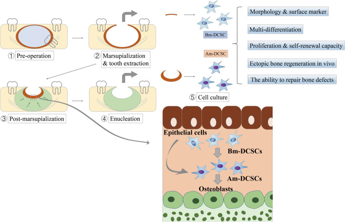
Dentigerous cyst (DC) is a bone destructive disease and remains a challenge for clinicians. Marsupialization enables the bone to regenerate with capsule maintaining, making it a preferred therapeutic means for DC adjacent to vital anatomical structures. Given that capsules of DC are derived from odontogenic epithelium remnants at the embryonic stage, we investigated whether there were mesenchymal stem cells (MSCs) located in DC capsules and the role that they played in the bone regeneration after marsupialization.

METHODS

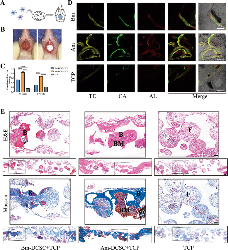
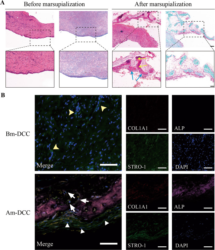
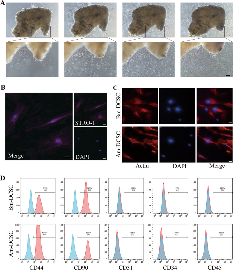
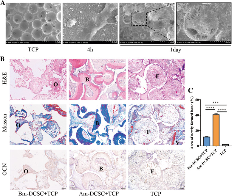
Samples obtained before and after marsupialization were used for histological detection and cell culture. The stemness of cells isolated from fresh tissues was analyzed by morphology, surface marker, and multi-differentiation assays. Comparison of proliferation ability between MSCs isolated from DC capsules before (Bm-DCSCs) and after (Am-DCSCs) marsupialization was evaluated by Cell Counting Kit-8 (CCK-8), fibroblast colony-forming units (CFU-F), and 5'-ethynyl-2'-deoxyuridine (EdU) assay. Their osteogenic capacity in vitro was detected by alkaline phosphatase (ALP) and Alizarin Red staining (ARS), combined with real-time polymerase chain reaction (RT-PCR) and immunofluorescence (IF) staining. Subcutaneous ectopic osteogenesis as well as cranial bone defect model in nude mice was performed to detect their bone regeneration and bone defect repairability.

RESULTS

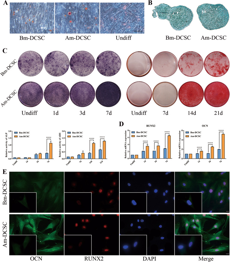
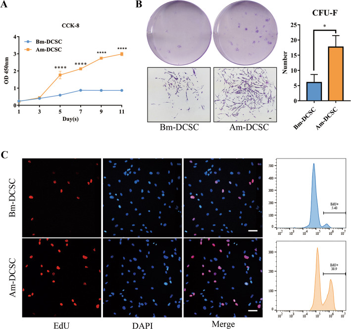
Bone tissue and strong ALP activity were detected in the capsule of DC after marsupialization. Two types of MSCs were isolated from fibrous capsules of DC both before (Bm-DCSCs) and after (Am-DCSCs) marsupialization. These fibroblast-like, colony-forming cells expressed MSC markers (CD44+, CD90+, CD31-, CD34-, CD45-), and they could differentiate into osteoblast-, adipocyte-, and chondrocyte-like cells under induction. Notably, Am-DCSCs performed better in cell proliferation and self-renewal. Moreover, Am-DCSCs showed a greater osteogenic capacity both in vitro and in vivo compared with Bm-DCSCs.

CONCLUSIONS

There are MSCs residing in capsules of DC, and the cell viability as well as the osteogenic capacity of them is largely enhanced after marsupialization. Our findings suggested that MSCs might play a crucial role in the healing process of DC after marsupialization, thus providing new insight into the treatment for DC by promoting the osteogenic differentiation of MSCs inside capsules.

摘要

背景

含牙囊肿(DC)是一种破坏骨骼的疾病,对临床医生来说仍是一个挑战。袋形术使骨骼在保持囊的情况下再生,使其成为毗邻重要解剖结构的 DC 的首选治疗手段。鉴于 DC 的囊源自胚胎期牙源性上皮的残余物,我们研究了在袋形术后 DC 囊内是否存在间充质干细胞(MSCs),以及它们在袋形术后骨再生中的作用。

方法

使用袋形术前和后的样本进行组织学检测和细胞培养。通过形态学、表面标志物和多分化试验分析从新鲜组织中分离的细胞的干性。通过细胞计数试剂盒-8(CCK-8)、成纤维细胞集落形成单位(CFU-F)和 5'-乙炔基-2'-脱氧尿苷(EdU)试验评估袋形术前(Bm-DCSCs)和术后(Am-DCSCs)分离的 MSCs 的增殖能力。通过碱性磷酸酶(ALP)和茜素红染色(ARS)检测其体外成骨能力,并结合实时聚合酶链反应(RT-PCR)和免疫荧光(IF)染色。在裸鼠中进行皮下异位成骨和颅骨缺损模型,以检测其骨再生和骨缺损修复能力。

结果

袋形术后 DC 囊内检测到骨组织和强 ALP 活性。从袋形术前(Bm-DCSCs)和术后(Am-DCSCs)的 DC 纤维囊中分离出两种类型的 MSC。这些成纤维细胞样、集落形成细胞表达 MSC 标志物(CD44+、CD90+、CD31-、CD34-、CD45-),并在诱导下可分化为成骨细胞、脂肪细胞和成软骨细胞样细胞。值得注意的是,Am-DCSCs 在细胞增殖和自我更新方面表现更好。此外,Am-DCSCs 在体外和体内均表现出比 Bm-DCSCs 更强的成骨能力。

结论

DC 囊内存在 MSCs,袋形术后其细胞活力和成骨能力大大增强。我们的研究结果表明,MSCs 可能在袋形术后 DC 的愈合过程中发挥关键作用,从而通过促进囊内 MSCs 的成骨分化,为 DC 的治疗提供新的思路。

https://cdn.ncbi.nlm.nih.gov/pmc/blobs/6ba4/7653780/43059d2fce3b/13287_2020_1999_Fig7_HTML.jpg
https://cdn.ncbi.nlm.nih.gov/pmc/blobs/6ba4/7653780/1565057daf1e/13287_2020_1999_Fig1_HTML.jpg
https://cdn.ncbi.nlm.nih.gov/pmc/blobs/6ba4/7653780/0533d3d9fb84/13287_2020_1999_Fig2_HTML.jpg
https://cdn.ncbi.nlm.nih.gov/pmc/blobs/6ba4/7653780/6a798a33f341/13287_2020_1999_Fig3_HTML.jpg
https://cdn.ncbi.nlm.nih.gov/pmc/blobs/6ba4/7653780/928b391932ac/13287_2020_1999_Fig4_HTML.jpg
https://cdn.ncbi.nlm.nih.gov/pmc/blobs/6ba4/7653780/100e586eaea2/13287_2020_1999_Fig5_HTML.jpg
https://cdn.ncbi.nlm.nih.gov/pmc/blobs/6ba4/7653780/15ad89dffc8c/13287_2020_1999_Fig6_HTML.jpg
https://cdn.ncbi.nlm.nih.gov/pmc/blobs/6ba4/7653780/43059d2fce3b/13287_2020_1999_Fig7_HTML.jpg

相似文献

1
Activation of mesenchymal stem cells promotes new bone formation within dentigerous cyst.间充质干细胞的激活促进含牙囊肿内新骨的形成。
Stem Cell Res Ther. 2020 Nov 10;11(1):476. doi: 10.1186/s13287-020-01999-8.
2
Co-culture of the bone and bone marrow: a novel way to obtain mesenchymal stem cells with enhanced osteogenic ability for fracture healing in SD rats.骨与骨髓共培养:一种提高骨髓间充质干细胞成骨能力促进 SD 大鼠骨折愈合的新方法。
J Orthop Surg Res. 2019 Sep 3;14(1):293. doi: 10.1186/s13018-019-1346-z.
3
MiR-374b promotes osteogenic differentiation of MSCs by degrading PTEN and promoting fracture healing.miR-374b 通过降解 PTEN 促进 MSC 的成骨分化,从而促进骨折愈合。
Eur Rev Med Pharmacol Sci. 2018 Jun;22(11):3303-3310. doi: 10.26355/eurrev_201806_15149.
4
MiR-140-5p promotes osteogenic differentiation of mouse embryonic bone marrow mesenchymal stem cells and post-fracture healing of mice.miR-140-5p 促进小鼠胚胎骨髓间充质干细胞的成骨分化和小鼠骨折后的愈合。
Cell Biochem Funct. 2020 Dec;38(8):1152-1160. doi: 10.1002/cbf.3585. Epub 2020 Oct 12.
5
Macrophages enhance mesenchymal stem cell osteogenesis via down-regulation of reactive oxygen species.巨噬细胞通过下调活性氧来增强间充质干细胞的成骨作用。
J Dent. 2020 Mar;94:103297. doi: 10.1016/j.jdent.2020.103297. Epub 2020 Feb 10.
6
MiR-125b Regulates the Osteogenic Differentiation of Human Mesenchymal Stem Cells by Targeting BMPR1b.微小RNA-125b通过靶向骨形态发生蛋白受体1b调控人间充质干细胞的成骨分化
Cell Physiol Biochem. 2017;41(2):530-542. doi: 10.1159/000457013. Epub 2017 Jan 31.
7
RIA fractions contain mesenchymal stroma cells with high osteogenic potency.放射免疫分析级分含有具有高成骨潜能的间充质基质细胞。
Injury. 2015 Dec;46 Suppl 8:S23-32. doi: 10.1016/S0020-1383(15)30051-6.
8
Mitochondria transfer enhances proliferation, migration, and osteogenic differentiation of bone marrow mesenchymal stem cell and promotes bone defect healing.线粒体转移增强骨髓间充质干细胞的增殖、迁移和成骨分化,并促进骨缺损愈合。
Stem Cell Res Ther. 2020 Jun 25;11(1):245. doi: 10.1186/s13287-020-01704-9.
9
Suppression of SPRY4 Promotes Osteogenic Differentiation and Bone Formation of Mesenchymal Stem Cell.抑制 SPRY4 促进间充质干细胞的成骨分化和骨形成。
Tissue Eng Part A. 2019 Dec;25(23-24):1646-1657. doi: 10.1089/ten.TEA.2019.0056. Epub 2019 Aug 9.
10
Homeobox C10 inhibits the osteogenic differentiation potential of mesenchymal stem cells.同源盒C10抑制间充质干细胞的成骨分化潜能。
Connect Tissue Res. 2018 May;59(3):201-211. doi: 10.1080/03008207.2017.1341496. Epub 2017 Aug 1.

引用本文的文献

1
Podoplanin immunoexpression in odontogenic lesions: a systematic review, meta-analysis, and integrated bioinformatic analysis.牙源性病变中 Podoplanin 的免疫表达:系统评价、荟萃分析和综合生物信息学分析。
Diagn Pathol. 2024 Aug 24;19(1):115. doi: 10.1186/s13000-024-01540-y.
2
Adult Mesenchymal Stem Cells from Oral Cavity and Surrounding Areas: Types and Biomedical Applications.来自口腔及周围区域的成人间充质干细胞:类型与生物医学应用
Pharmaceutics. 2023 Aug 9;15(8):2109. doi: 10.3390/pharmaceutics15082109.
3
miR-30a inhibits the osteogenic differentiation of the tibia-derived MSCs in congenital pseudarthrosis via targeting HOXD8.

本文引用的文献

1
Guidelines and definitions for research on epithelial-mesenchymal transition.上皮-间质转化研究的指南和定义。
Nat Rev Mol Cell Biol. 2020 Jun;21(6):341-352. doi: 10.1038/s41580-020-0237-9. Epub 2020 Apr 16.
2
Five Years Follow-up of a Spontaneous Eruption of an Impacted Mandibular Premolar Associated with a Dentigerous Cyst Treated by Marsupialization.对通过袋形术治疗的与含牙囊肿相关的下颌阻生前磨牙自然萌出进行的五年随访
Cureus. 2020 Mar 22;12(3):e7370. doi: 10.7759/cureus.7370.
3
The osteocyte: A multifunctional cell within the bone.
微小RNA-30a通过靶向同源盒D8抑制先天性假关节中胫骨来源间充质干细胞的成骨分化。
Regen Ther. 2022 Oct 20;21:477-485. doi: 10.1016/j.reth.2022.09.005. eCollection 2022 Dec.
成骨细胞:骨骼中的多功能细胞。
Ann Anat. 2020 Jan;227:151422. doi: 10.1016/j.aanat.2019.151422. Epub 2019 Sep 26.
4
Marsupialization of a dentigerous cyst followed by orthodontic traction of two retained teeth: A case report.含牙囊肿袋形术联合两颗滞留牙的正畸牵引:一例报告
Int Orthod. 2019 Jun;17(2):365-374. doi: 10.1016/j.ortho.2019.03.019. Epub 2019 Apr 22.
5
Marsupialization of a Large Radicular Cyst with Extensive Maxillary Tooth Displacement: Eight-Year Follow-Up.伴有广泛上颌牙移位的大型根端囊肿袋形术:八年随访
J Dent Child (Chic). 2019 Jan 15;86(1):64-68.
6
Cyst volume changes measured with a 3D reconstruction after decompression of a mandibular dentigerous cyst with an impacted third molar.在下颌含牙囊肿减压并拔除阻生第三磨牙后,通过三维重建测量囊肿体积变化。
Clin Pract. 2019 Feb 26;9(1):1132. doi: 10.4081/cp.2019.1132. eCollection 2019 Jan 29.
7
Expression of keratin 15 in dentigerous cyst, odontogenic keratocyst and ameloblastoma.角蛋白15在含牙囊肿、牙源性角化囊肿和成釉细胞瘤中的表达。
Mol Clin Oncol. 2019 Mar;10(3):377-381. doi: 10.3892/mco.2019.1802. Epub 2019 Jan 17.
8
Commitment of Oral-Derived Stem Cells in Dental and Maxillofacial Applications.口腔源性干细胞在口腔颌面应用中的定向分化
Dent J (Basel). 2018 Dec 13;6(4):72. doi: 10.3390/dj6040072.
9
Recurrence of nonsyndromic odontogenic keratocyst after marsupialization and delayed enucleation vs. enucleation alone: a systematic review and meta-analysis.袋形术联合延期摘除术与单纯摘除术治疗非综合征性牙源性角化囊肿后的复发情况:一项系统评价和荟萃分析
Oral Maxillofac Surg. 2019 Mar;23(1):1-11. doi: 10.1007/s10006-018-0737-3. Epub 2018 Nov 29.
10
Concise Review: Cellular and Molecular Mechanisms Regulation of Tooth Initiation.简明综述:牙齿起始的细胞和分子机制调控。
Stem Cells. 2019 Jan;37(1):26-32. doi: 10.1002/stem.2917. Epub 2018 Nov 20.