Hong Jun-Jie, Liu Bo-le, Wang Zhi-Qiang, Tang Kun, Ji Xiao-Wei, Yin Wei-Wei, Lin Jie, Zheng Xiang-Wu
Department of PET/CT, Radiology Imaging Center, The First Affiliated Hospital of Wenzhou Medical University, Xuefu North Rd, Wenzhou, 325000, Zhejiang, People's Republic of China.
EJNMMI Res. 2020 Nov 10;10(1):138. doi: 10.1186/s13550-020-00730-1.
Clinical management decisions on prostate cancer (PCa) are often based on a determination of risk. Ga-prostate-specific membrane antigen (PSMA)-11-positron emission tomography (PET)/computer tomography (CT) is an attractive modality to assess biochemical recurrence of PCa, detect metastatic disease and stage of primary PCa, making it a promising strategy for risk stratification. However, due to some limitation of Ga-PSMA-11 the development of alternative tracers is of high interest. In this study, we aimed to investigate the value of F-PSMA-1007 in identifying non-metastatic high-risk PCa.
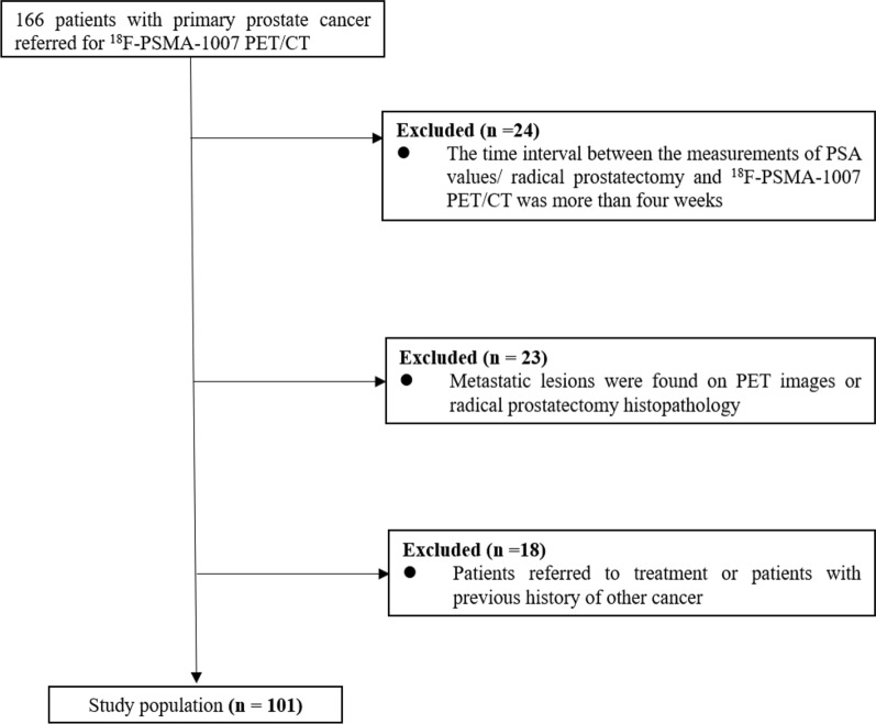
A total of 101 patients with primary non-metastatic PCa who underwent F-PSMA-1007 PET/CT were retrospectively analyzed. According to the European Association of Urology guidelines on PCa, patients were classified into intermediate-risk (IR) group or high-risk (HR) group. The maximum standardized uptake values (SUVmax) of the primary prostate tumor were measured on PET/CT images. The diagnostic performance of PET/CT for IR and HR PCa was calculated, and the relationship between the SUVmax of primary prostate tumor, prostate-specific antigen (PSA) level and Gleason score (GS) was analyzed.
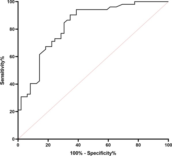
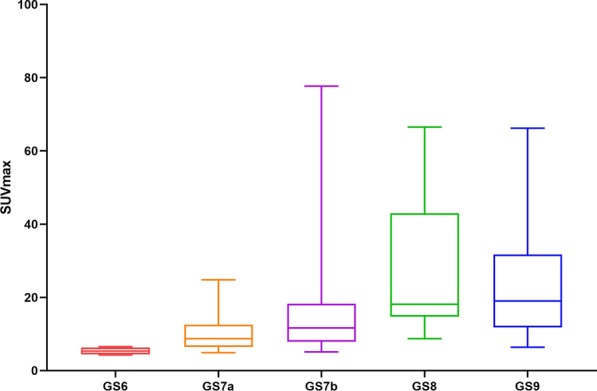
Of all 101 patients, 49 patients were classified into IR group and 52 patients were classified into HR group. There was a significant positive correlation between PSA level/GS and SUVmax (r = 0.561, r = 0.496, P < 0.001, respectively). Tumors with GS 6 and 7a showed significantly lower F-PSMA-1007 uptake compared to patients with GS 8 and 9 (P < 0.01). SUVmax in patients of HR was significantly higher than those of IR (median SUVmax: 16.85 vs 7.80; P < 0.001). In receiver operating characteristic curve analysis, the optimal cutoff value of the SUVmax for identifying high-risk PCa was set as 9.05 (area under the curve: 0.829; sensitivity: 90.4%; specificity: 65.3%).
F-PSMA-1007 PET/CT showed the powerful diagnosis efficacy for high-risk PCa, which can be used as an objective imaging reference index for clinical reference.
前列腺癌(PCa)的临床管理决策通常基于风险评估。镓-前列腺特异性膜抗原(PSMA)-11正电子发射断层扫描(PET)/计算机断层扫描(CT)是评估PCa生化复发、检测转移性疾病和原发性PCa分期的一种有吸引力的方法,使其成为一种有前景的风险分层策略。然而,由于镓-PSMA-11存在一些局限性,开发替代示踪剂备受关注。在本研究中,我们旨在探讨F-PSMA-1007在识别非转移性高危PCa中的价值。
回顾性分析了101例接受F-PSMA-1007 PET/CT检查的原发性非转移性PCa患者。根据欧洲泌尿外科协会关于PCa的指南,将患者分为中危(IR)组或高危(HR)组。在PET/CT图像上测量原发性前列腺肿瘤的最大标准化摄取值(SUVmax)。计算PET/CT对IR和HR PCa的诊断性能,并分析原发性前列腺肿瘤的SUVmax、前列腺特异性抗原(PSA)水平和 Gleason评分(GS)之间的关系。
101例患者中,49例患者被分为IR组,52例患者被分为HR组。PSA水平/GS与SUVmax之间存在显著正相关(r分别为0.561和0.496,P<0.001)。与GS 8和9的患者相比,GS 6和7a的肿瘤显示出显著更低的F-PSMA-1007摄取(P<0.01)。HR组患者的SUVmax显著高于IR组(SUVmax中位数:16.85对7.80;P<0.001)。在受试者工作特征曲线分析中,用于识别高危PCa的SUVmax的最佳截断值设定为9.05(曲线下面积:0.829;灵敏度:90.4%;特异性:65.3%)。
F-PSMA-1007 PET/CT对高危PCa显示出强大的诊断效能,可作为临床参考的客观影像指标。