Tanaka Shoichiro A, Patel Nilesh M, Murthy Ananth S
Akron Children's Hospital, Akron, Ohio.
Michigan Spine Clinic, Brownstown, Mich.
Plast Reconstr Surg Glob Open. 2020 Oct 29;8(10):e3196. doi: 10.1097/GOX.0000000000003196. eCollection 2020 Oct.
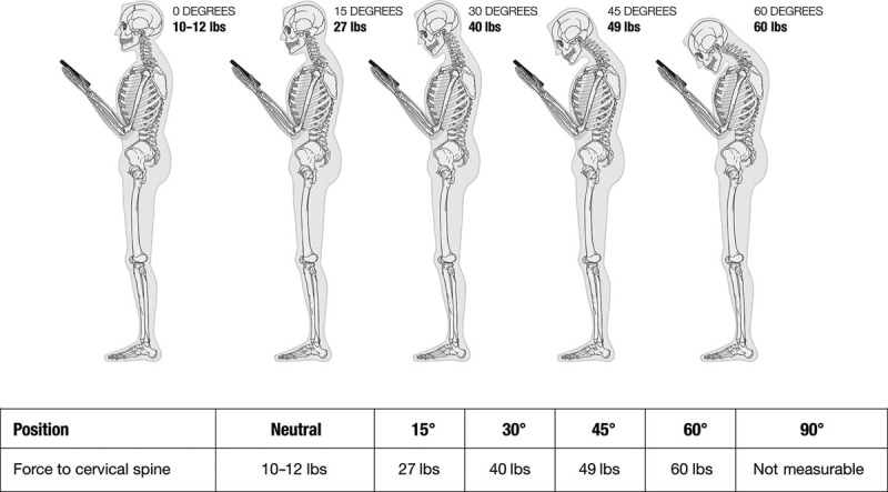
For craniofacial surgeons, cleft palate repair is an intricate and difficult operation positionally. Historically, use of loupe magnification and a headlight can cause significant strain to the surgeon's neck and, at times, subpar optics for both the operator and the assistant. The use of an operating microscope was first advocated by Sommerlad in 2003. By using the operating microscope for cleft palate closure, there are improved ergonomics for the surgeon and assistant by allowing for straight in-line back and neck posture with excellent visualization of the surgical field for the entire surgical team. The available zoom and focus improve the ability to isolate and repair the levator veli palatini muscle. Proper posture with a neutral cervical spine will help prolong a surgeon's career and ability to care for their patients.
对于颅面外科医生来说,腭裂修复手术在操作位置上是一项复杂且困难的手术。从历史上看,使用头戴式放大镜和头灯会给外科医生的颈部带来很大压力,而且有时对于术者和助手来说光学效果都不尽人意。手术显微镜的使用最早是由索默拉德在2003年倡导的。通过使用手术显微镜进行腭裂修复,外科医生和助手的人体工程学得到了改善,因为可以保持背部和颈部挺直的直线姿势,整个手术团队都能很好地观察手术区域。可用的变焦和聚焦功能提高了分离和修复腭帆提肌的能力。保持颈椎中立的正确姿势将有助于延长外科医生的职业生涯以及照顾患者的能力。