Department of Diagnostic and Interventional Radiology, Faculty of Medicine and University Hospital Cologne, University of Cologne, Kerpener Str. 62, 50937, Cologne, Germany.
Department of Diagnostic and Interventional Radiology, University Düsseldorf, Medical Faculty, 40225, Düsseldorf, Germany.
Eur Radiol. 2021 May;31(5):3468-3477. doi: 10.1007/s00330-020-07379-3. Epub 2020 Nov 12.
To investigate whether the increased soft tissue contrast of virtual monoenergetic images (VMIs) obtained from a spectral detector computed tomography (SDCT) system improves washout assessment of arterially hyper-enhancing liver lesions.
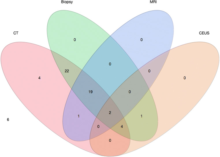
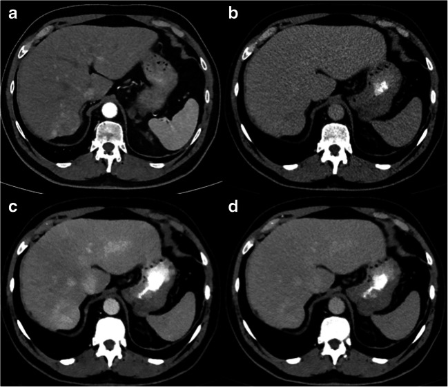
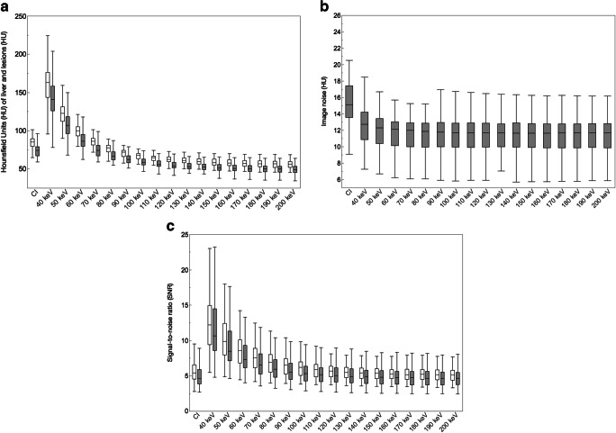
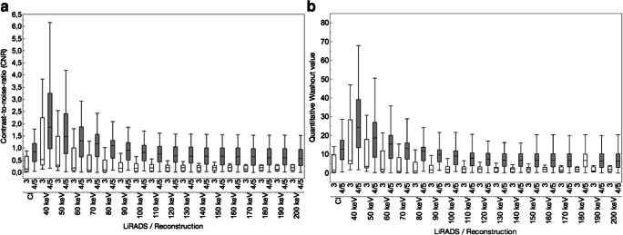
Fifty-nine arterially hyper-enhancing lesions in 31 patients (age 65 ± 9 years, M/W 20/11) were included in this IRB-approved study. All patients underwent multi-phase SDCT for HCC screening. MRI, CEUS or biopsy within 3 months served as standard of reference to classify lesions as LiRADS 3 or 4/5. VMIs and conventional images (CIs) were reconstructed. Visual analysis was performed on 40, 60, and 80 kiloelectronvolt (keV) and CIs by 3 radiologists. Presence and visibility of washout were assessed; image quality and confidence of washout evaluation were evaluated on 5-point Likert scales. Signal-to-noise ratio (SNR), lesion-to-liver contrast-to-noise ratio (CNR) (|HU-HU|/SD) and washout (|HU-HU|) were calculated. Statistical assessment was performed using ANOVA and Wilcoxon test.
On subjective lesion analysis, the highest level of diagnostic confidence and highest sensitivity for the detection of lesion washout were found for 40-keV VMIs (40 keV vs. CI, 81.3 vs. 71.3%). Image quality parameters were significantly better in low-kiloelectronvolt VMIs than in CIs (p < 0.05; e.g. SNR: 40 keV vs. CIs, 12.5 ± 4.1 vs. 5.6 ± 1.6). In LiRADS 4/5 lesions, CNR and quantitative washout values were significantly higher in 40-keV VMIs compared to CIs (p < 0.05; e.g. CNR and washout in 40 keV vs. CIs, 2.3 ± 1.6 vs. 0.8 ± 0.5 and 29.0 ± 19.1 vs. 12.9 ± 6.9 HU, respectively).
By increasing lesion contrast, low-kiloelectronvolt VMIs obtained from SDCT improve washout assessment of hyper-enhancing liver lesions with respect to washout visibility and diagnostic confidence.
• Low-kiloelectronvolt virtual monoenergetic images from spectral detector CT facilitate washout assessment in arterially hyper-enhancing liver lesions. • Image quality and quantitative washout parameters as well as subjective washout visibility and diagnostic confidence benefit from low-kiloelectronvolt virtual monoenergetic images.
探讨能谱 CT 系统得到的软组织对比度增加的虚拟单能量图像(VMIs)是否改善动脉期高增强肝病变的洗脱评估。
本研究经机构审查委员会批准,纳入 31 例患者的 59 个动脉期高增强病变(年龄 65±9 岁,男/女 20/11)。所有患者均行多期能谱 CT 进行 HCC 筛查。MRI、CEUS 或 3 个月内的活检作为分类病变为 LiRADS 3 或 4/5 的标准。重建 VMIs 和常规图像(CIs)。3 名放射科医生对 40、60 和 80keV 的 VMIs 和 CIs 进行视觉分析。评估洗脱的存在和可视性;对图像质量和洗脱评估的置信度进行 5 分李克特量表评估。计算信噪比(SNR)、病变与肝脏对比噪声比(CNR)[|HU-HU|/SD]和洗脱(|HU-HU|)。使用方差分析和 Wilcoxon 检验进行统计学评估。
在主观病变分析中,40keV VMIs 的诊断信心水平最高,检测病变洗脱的灵敏度最高(40keV 比 CI,81.3%比 71.3%)。低千伏 VMIs 的图像质量参数明显优于 CIs(p<0.05;例如 SNR:40keV 比 CIs,12.5±4.1 比 5.6±1.6)。在 LiRADS 4/5 病变中,与 CIs 相比,40keV VMIs 的 CNR 和定量洗脱值明显更高(p<0.05;例如 CNR 和洗脱在 40keV 比 CIs,2.3±1.6 比 0.8±0.5 和 29.0±19.1 比 12.9±6.9HU)。
通过增加病变对比度,能谱 CT 得到的低千伏 VMIs 可改善动脉期高增强肝脏病变的洗脱评估,包括洗脱可见度和诊断信心。
• 光谱探测器 CT 的低千伏虚拟单能量图像有助于动脉期高增强肝脏病变的洗脱评估。• 图像质量和定量洗脱参数以及主观洗脱可见度和诊断信心受益于低千伏虚拟单能量图像。