Weigele Jochen, Bohnsack Brenda L
Division of Ophthalmology, Ann & Robert H. Lurie Children's Hospital of Chicago, 225 E. Chicago Ave, Chicago, IL 60611, USA.
Department of Ophthalmology, Northwestern University Feinberg School of Medicine, 645 N. Michigan Ave, Chicago, IL 60611, USA.
J Dev Biol. 2020 Nov 10;8(4):26. doi: 10.3390/jdb8040026.
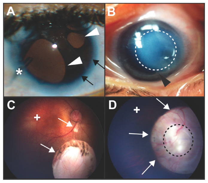
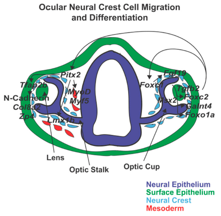
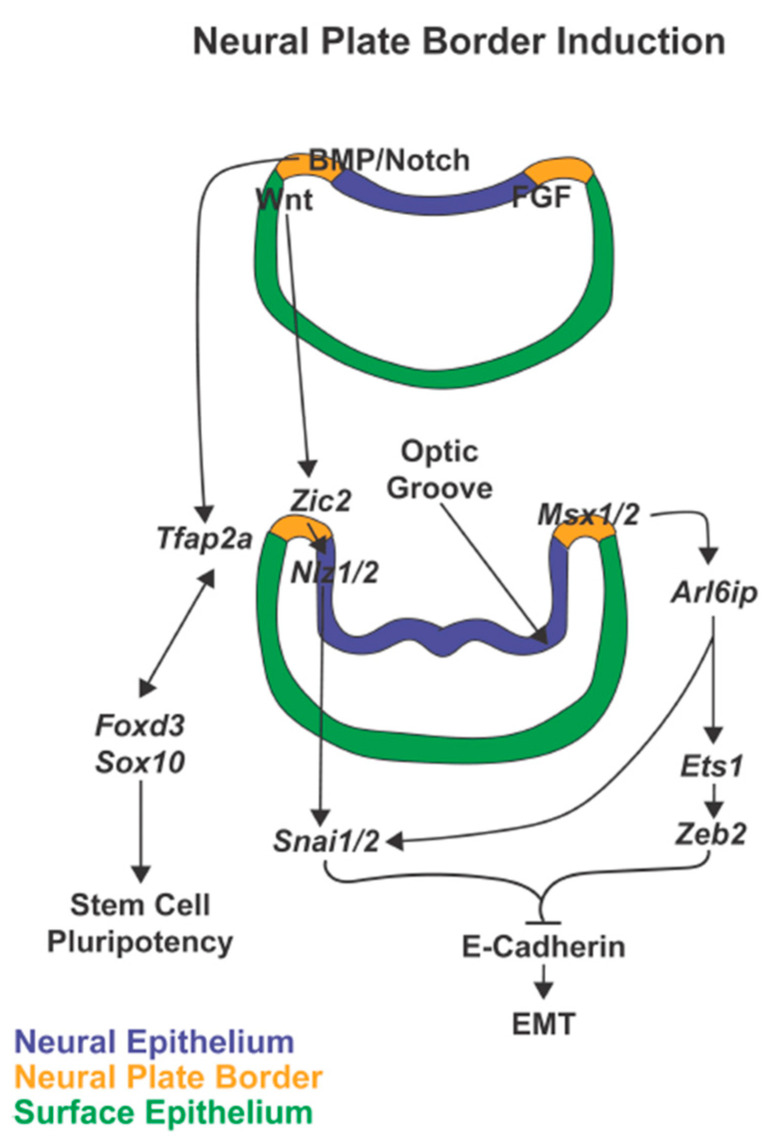
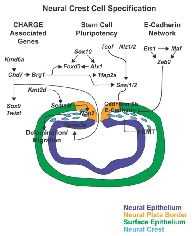
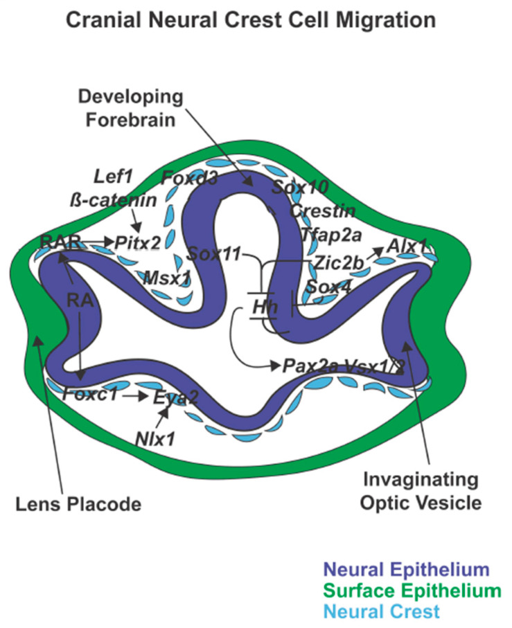
The neural crest is a unique, transient stem cell population that is critical for craniofacial and ocular development. Understanding the genetics underlying the steps of neural crest development is essential for gaining insight into the pathogenesis of congenital eye diseases. The neural crest cells play an under-appreciated key role in patterning the neural epithelial-derived optic cup. These interactions between neural crest cells within the periocular mesenchyme and the optic cup, while not well-studied, are critical for optic cup morphogenesis and ocular fissure closure. As a result, microphthalmia and coloboma are common phenotypes in human disease and animal models in which neural crest cell specification and early migration are disrupted. In addition, neural crest cells directly contribute to numerous ocular structures including the cornea, iris, sclera, ciliary body, trabecular meshwork, and aqueous outflow tracts. Defects in later neural crest cell migration and differentiation cause a constellation of well-recognized ocular anterior segment anomalies such as Axenfeld-Rieger Syndrome and Peters Anomaly. This review will focus on the genetics of the neural crest cells within the context of how these complex processes specifically affect overall ocular development and can lead to congenital eye diseases.
神经嵴是一种独特的、短暂存在的干细胞群体,对颅面和眼部发育至关重要。了解神经嵴发育步骤背后的遗传学对于深入了解先天性眼病的发病机制至关重要。神经嵴细胞在构建神经上皮来源的视杯过程中发挥着未得到充分重视的关键作用。眼周间充质内的神经嵴细胞与视杯之间的这些相互作用,虽然研究尚不充分,但对视杯形态发生和眼裂闭合至关重要。因此,小眼症和脉络膜缺损是人类疾病和动物模型中的常见表型,其中神经嵴细胞的特化和早期迁移受到破坏。此外,神经嵴细胞直接参与形成许多眼部结构,包括角膜、虹膜、巩膜、睫状体、小梁网和房水流出通道。后期神经嵴细胞迁移和分化的缺陷会导致一系列公认的眼前段异常,如Axenfeld-Rieger综合征和彼得斯异常。本综述将聚焦于神经嵴细胞的遗传学,探讨这些复杂过程如何具体影响整体眼部发育并导致先天性眼病。