Department of Gastroenterology and Hepatology, University Medical Center Groningen, University of Groningen, 9712 CP Groningen, The Netherlands.
Department of Pediatrics, University Medical Center Groningen, University of Groningen, 9712 CP Groningen, The Netherlands.
Cells. 2020 Nov 11;9(11):2456. doi: 10.3390/cells9112456.
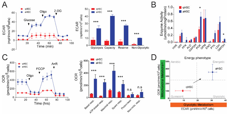
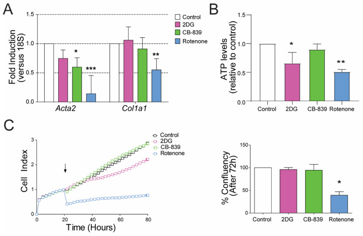
Upon liver injury, hepatic stellate cells (HSCs) transdifferentiate to migratory, proliferative and extracellular matrix-producing myofibroblasts (e.g., activated HSCs; aHSCs) causing liver fibrosis. HSC activation is associated with increased glycolysis and glutaminolysis. Here, we compared the contribution of glycolysis, glutaminolysis and mitochondrial oxidative phosphorylation (OXPHOS) in rat and human HSC activation. Basal levels of glycolysis (extracellular acidification rate ~3-fold higher) and particularly mitochondrial respiration (oxygen consumption rate ~5-fold higher) were significantly increased in rat aHSCs, when compared to quiescent rat HSC. This was accompanied by extensive mitochondrial fusion in rat and human aHSCs, which occurred without increasing mitochondrial DNA content and electron transport chain (ETC) components. Inhibition of glycolysis (by 2-deoxy-D-glucose) and glutaminolysis (by CB-839) did not inhibit rat aHSC proliferation, but did reduce (encoding α-SMA) expression slightly. In contrast, inhibiting mitochondrial OXPHOS (by rotenone) significantly suppressed rat aHSC proliferation, as well as and expression. Other than that observed for rat aHSCs, human aHSC proliferation and expression of fibrosis markers were significantly suppressed by inhibiting either glycolysis, glutaminolysis or mitochondrial OXPHOS (by metformin). Activation of HSCs is marked by simultaneous induction of glycolysis and mitochondrial metabolism, extending the possibilities to suppress hepatic fibrogenesis by interfering with HSC metabolism.
在肝损伤时,肝星状细胞(HSCs)向迁移、增殖和细胞外基质产生的肌成纤维细胞(例如,活化的 HSCs;aHSCs)分化,导致肝纤维化。HSC 的活化与糖酵解和谷氨酰胺分解的增加有关。在这里,我们比较了糖酵解、谷氨酰胺分解和线粒体氧化磷酸化(OXPHOS)在大鼠和人 HSC 活化中的作用。与静止的大鼠 HSCs 相比,大鼠 aHSCs 的基础糖酵解水平(细胞外酸化率高约 3 倍)和特别是线粒体呼吸(耗氧率高约 5 倍)显著增加。这伴随着大鼠和人 aHSCs 中广泛的线粒体融合,而线粒体融合并没有增加线粒体 DNA 含量和电子传递链(ETC)成分。抑制糖酵解(通过 2-脱氧-D-葡萄糖)和谷氨酰胺分解(通过 CB-839)并没有抑制大鼠 aHSC 的增殖,但确实略微降低了 (编码 α-SMA)的表达。相比之下,抑制线粒体 OXPHOS(通过鱼藤酮)显著抑制了大鼠 aHSC 的增殖以及 和 的表达。除了在大鼠 aHSCs 中观察到的情况外,通过抑制糖酵解、谷氨酰胺分解或线粒体 OXPHOS(通过二甲双胍),人 aHSC 的增殖和纤维化标志物的表达也明显受到抑制。HSCs 的活化伴随着糖酵解和线粒体代谢的同时诱导,这为通过干扰 HSC 代谢来抑制肝纤维化提供了更多的可能性。