• 文献检索
  • 文档翻译
  • 深度研究
  • 学术资讯
  • Suppr Zotero 插件Zotero 插件
  • 邀请有礼
  • 套餐&价格
  • 历史记录
应用&插件
Suppr Zotero 插件Zotero 插件浏览器插件Mac 客户端Windows 客户端微信小程序
定价
高级版会员购买积分包购买API积分包
服务
文献检索文档翻译深度研究API 文档MCP 服务
关于我们
关于 Suppr公司介绍联系我们用户协议隐私条款
关注我们

Suppr 超能文献

核心技术专利:CN118964589B侵权必究
粤ICP备2023148730 号-1Suppr @ 2026

文献检索

告别复杂PubMed语法,用中文像聊天一样搜索,搜遍4000万医学文献。AI智能推荐,让科研检索更轻松。

立即免费搜索

文件翻译

保留排版,准确专业,支持PDF/Word/PPT等文件格式,支持 12+语言互译。

免费翻译文档

深度研究

AI帮你快速写综述,25分钟生成高质量综述,智能提取关键信息,辅助科研写作。

立即免费体验

组蛋白修饰分析揭示小鼠模型腹主动脉瘤形成过程中的表观基因组动态变化。

Profiling of Histone Modifications Reveals Epigenomic Dynamics During Abdominal Aortic Aneurysm Formation in Mouse Models.

作者信息

Greenway Jacob, Gilreath Nicole, Patel Sagar, Horimatsu Tetsuo, Moses Mary, Kim David, Reid Lauren, Ogbi Mourad, Shi Yang, Lu Xin-Yun, Shukla Mrinal, Lee Richard, Huo Yuqing, Young Lufei, Kim Ha Won, Weintraub Neal L

机构信息

Departments of Medicine, College of Nursing at Augusta University, Augusta, GA, United States.

Vascular Biology Center, Medical College of Georgia, College of Nursing at Augusta University, Augusta, GA, United States.

出版信息

Front Cardiovasc Med. 2020 Oct 30;7:595011. doi: 10.3389/fcvm.2020.595011. eCollection 2020.

DOI:10.3389/fcvm.2020.595011
PMID:33195484
原文链接:https://pmc.ncbi.nlm.nih.gov/articles/PMC7662126/
Abstract

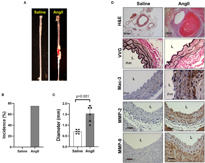
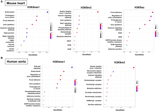
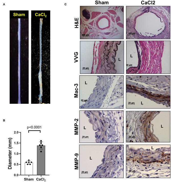
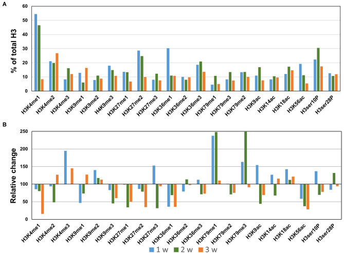
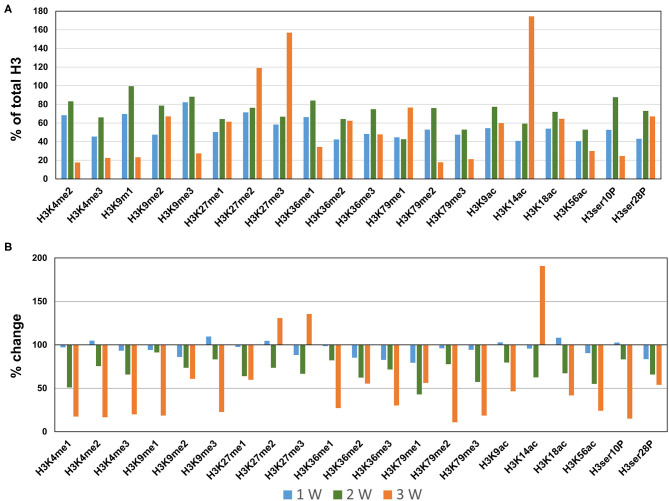
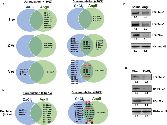
Abdominal aortic aneurysms (AAA) are characterized by localized inflammation, extracellular matrix degradation, and apoptosis of smooth muscle cells, which together lead to progressive and irreversible aortic dilation. Major risk factors for AAA include smoking and aging, both of which prominently alter gene expression via epigenetic mechanisms, including histone methylation (me) and acetylation (ac).However, little is known about epigenomic dynamics during AAA formation. Here, we profiled histone modification patterns in aortic tissues during AAA formation in two distinct mouse models; (1) angiotensin II (AngII) infusion in low density lipoprotein receptor (LDLR) knockout (KO) mice, and (2) calcium chloride (CaCl) application in wild type mice. AAA formed in both models, in conjunction with enhanced macrophage infiltration, elastin degradation and matrix metalloproteinases expression as evaluated by immunohistochemistry. To investigate the histone modification patterns during AAA formation, total histone proteins were extracted from AAA tissues, and histone H3 modifications were quantified using profiling kits. Intriguingly, we observed dynamic changes in histone H3 modifications of lysine (K) residues at different time points during AAA formation. In mature aneurysmal tissues at 3 weeks after AngII infusion, we detected reduced K4/K27/K36 monomethylation, K9 trimethylation K9, and K9/K56 acetylation (<70%), and increased K4 trimethylation (>130%). Conversely, in CaCl-induced AAA, K4/K9/K27/K36/K79 monomethylation and K9/K18/K56 acetylation were reduced in AAA tissues, whereas K27 di-/tri-methylation and K14 acetylation were upregulated. Interestingly, K4/K27/K36 monomethylation, K9 trimethylation, and K9/K56 acetylation were commonly downregulated in both animal models, while no H3 modifications were uniformly upregulated. Western blot of AAA tissues confirmed markedly reduced levels of key H3 modifications, including H3K4me1, H3K9me3, and H3K56ac. Furthermore, pathway enrichment analysis using an integrative bioinformatics approach identified specific molecular pathways, including endocytosis, exon guidance and focal adhesion signaling, that may potentially be linked to these histone H3 modifications during AAA formation. Dynamic modifications of histone H3 occur during AAA formation in both animal models. We identified 6 discreet H3 modifications that are consistently downregulated in both models, suggesting a possible role in AAA pathobiology. Identifying the functional mechanisms may facilitate development of novel strategies for AAA prevention or treatment.

摘要

腹主动脉瘤(AAA)的特征是局部炎症、细胞外基质降解和平滑肌细胞凋亡,这些共同导致主动脉进行性和不可逆的扩张。AAA的主要危险因素包括吸烟和衰老,这两者均通过表观遗传机制显著改变基因表达,包括组蛋白甲基化(me)和乙酰化(ac)。然而,关于AAA形成过程中的表观基因组动态变化知之甚少。在此,我们在两种不同的小鼠模型中分析了AAA形成过程中主动脉组织中的组蛋白修饰模式;(1)在低密度脂蛋白受体(LDLR)基因敲除(KO)小鼠中输注血管紧张素II(AngII),以及(2)在野生型小鼠中应用氯化钙(CaCl)。两种模型中均形成了AAA,免疫组织化学评估显示巨噬细胞浸润增强、弹性蛋白降解和基质金属蛋白酶表达增加。为了研究AAA形成过程中的组蛋白修饰模式,从AAA组织中提取总组蛋白,并使用分析试剂盒对组蛋白H3修饰进行定量。有趣的是,我们观察到AAA形成过程中不同时间点赖氨酸(K)残基的组蛋白H3修饰发生了动态变化。在AngII输注后3周的成熟动脉瘤组织中,我们检测到K4/K27/K36单甲基化、K9三甲基化K9和K9/K56乙酰化降低(<70%),以及K4三甲基化增加(>130%)。相反,在CaCl诱导的AAA中,AAA组织中K4/K9/K27/K36/K79单甲基化和K9/K18/K56乙酰化降低,而K27二/三甲基化和K14乙酰化上调。有趣地是,在两种动物模型中,K4/K27/K36单甲基化、K9三甲基化和K9/K56乙酰化均普遍下调,而没有H3修饰被一致上调。AAA组织的蛋白质印迹证实关键H3修饰水平显著降低,包括H3K4me1、H3K9me3和H3K56ac。此外,使用综合生物信息学方法进行的通路富集分析确定了特定的分子通路,包括内吞作用、外显子引导和粘着斑信号传导,这些通路可能在AAA形成过程中与这些组蛋白H3修饰潜在相关。在两种动物模型的AAA形成过程中均发生了组蛋白H3的动态修饰。我们确定了6种在两种模型中均持续下调的离散H3修饰,表明其在AAA病理生物学中可能发挥作用。确定其功能机制可能有助于开发预防或治疗AAA的新策略。

https://cdn.ncbi.nlm.nih.gov/pmc/blobs/6d2f/7662126/18b7681e3230/fcvm-07-595011-g0006.jpg
https://cdn.ncbi.nlm.nih.gov/pmc/blobs/6d2f/7662126/f357f882961d/fcvm-07-595011-g0001.jpg
https://cdn.ncbi.nlm.nih.gov/pmc/blobs/6d2f/7662126/6f7f28e73bff/fcvm-07-595011-g0002.jpg
https://cdn.ncbi.nlm.nih.gov/pmc/blobs/6d2f/7662126/174d2700a60d/fcvm-07-595011-g0003.jpg
https://cdn.ncbi.nlm.nih.gov/pmc/blobs/6d2f/7662126/77069bd163ce/fcvm-07-595011-g0004.jpg
https://cdn.ncbi.nlm.nih.gov/pmc/blobs/6d2f/7662126/9c496147bc8e/fcvm-07-595011-g0005.jpg
https://cdn.ncbi.nlm.nih.gov/pmc/blobs/6d2f/7662126/18b7681e3230/fcvm-07-595011-g0006.jpg

相似文献

1
Profiling of Histone Modifications Reveals Epigenomic Dynamics During Abdominal Aortic Aneurysm Formation in Mouse Models.组蛋白修饰分析揭示小鼠模型腹主动脉瘤形成过程中的表观基因组动态变化。
Front Cardiovasc Med. 2020 Oct 30;7:595011. doi: 10.3389/fcvm.2020.595011. eCollection 2020.
2
Distinct patterns of histone methylation and acetylation in human interphase nuclei.人类间期细胞核中组蛋白甲基化和乙酰化的独特模式。
Physiol Res. 2007;56(6):797-806. doi: 10.33549/physiolres.931123. Epub 2007 Feb 8.
3
Structural cooperativity in histone H3 tail modifications.组蛋白 H3 尾部修饰的结构协同性。
Protein Sci. 2011 Dec;20(12):1982-90. doi: 10.1002/pro.745. Epub 2011 Oct 19.
4
[The roles of histone lysine methylation in epigenetic regulation].[组蛋白赖氨酸甲基化在表观遗传调控中的作用]
Yi Chuan. 2007 Apr;29(4):387-92. doi: 10.1360/yc-007-0387.
5
Promoter histone H3 lysine 9 di-methylation is associated with DNA methylation and aberrant expression of p16 in gastric cancer cells.启动子组蛋白H3赖氨酸9二甲基化与胃癌细胞中的DNA甲基化及p16异常表达相关。
Oncol Rep. 2009 Nov;22(5):1221-7. doi: 10.3892/or_00000558.
6
The organization of histone H3 modifications as revealed by a panel of specific monoclonal antibodies.一组特异性单克隆抗体所揭示的组蛋白H3修饰的组织方式。
Cell Struct Funct. 2008;33(1):61-73. doi: 10.1247/csf.07035. Epub 2008 Jan 28.
7
Histone H3 acetylation and H3 K4 methylation define distinct chromatin regions permissive for transgene expression.组蛋白H3乙酰化和H3 K4甲基化定义了允许转基因表达的不同染色质区域。
Mol Cell Biol. 2006 Sep;26(17):6357-71. doi: 10.1128/MCB.00311-06.
8
Spatial distribution of di- and tri-methyl lysine 36 of histone H3 at active genes.活性基因处组蛋白H3的二甲基赖氨酸36和三甲基赖氨酸36的空间分布。
J Biol Chem. 2005 May 6;280(18):17732-6. doi: 10.1074/jbc.M500796200. Epub 2005 Mar 10.
9
Histone H3 Acetyl K9 and Histone H3 Tri Methyl K4 as Prognostic Markers for Patients with Cervical Cancer.组蛋白H3乙酰化赖氨酸9和组蛋白H3三甲基化赖氨酸4作为宫颈癌患者的预后标志物
Int J Mol Sci. 2017 Feb 23;18(3):477. doi: 10.3390/ijms18030477.
10
β-Carotene enhances the expression of inflammation-related genes and histone H3 K9 acetylation, K4 dimethylation, and K36 trimethylation around these genes in juvenile macrophage-like THP-1 cells.β-胡萝卜素可增强幼年巨噬细胞样THP-1细胞中炎症相关基因的表达以及这些基因周围的组蛋白H3 K9乙酰化、K4二甲基化和K36三甲基化。
Biochem Biophys Rep. 2022 Aug 15;31:101325. doi: 10.1016/j.bbrep.2022.101325. eCollection 2022 Sep.

引用本文的文献

1
Microinjection of angiotensin II into zebrafish embryos induces transient dilation and elastin disruption of the dorsal aorta.向斑马鱼胚胎显微注射血管紧张素II会诱导背主动脉短暂扩张和弹性蛋白破坏。
Physiol Rep. 2025 Feb;13(4):e70259. doi: 10.14814/phy2.70259.
2
Epigenetic modifications in abdominal aortic aneurysms: from basic to clinical.腹主动脉瘤中的表观遗传修饰:从基础到临床
Front Cardiovasc Med. 2024 Jun 4;11:1394889. doi: 10.3389/fcvm.2024.1394889. eCollection 2024.
3
Adenosine kinase inhibition protects mice from abdominal aortic aneurysm via epigenetic modulation of VSMC inflammation.

本文引用的文献

1
Niacin protects against abdominal aortic aneurysm formation via GPR109A independent mechanisms: role of NAD+/nicotinamide.烟酸通过 GPR109A 非依赖机制预防腹主动脉瘤形成:NAD+/烟酰胺的作用。
Cardiovasc Res. 2020 Dec 1;116(14):2226-2238. doi: 10.1093/cvr/cvz303.
2
Epigenetic regulation of regulatory T cells in patients with abdominal aortic aneurysm.腹主动脉瘤患者调节性 T 细胞的表观遗传调控。
FEBS Open Bio. 2019 Jun;9(6):1137-1143. doi: 10.1002/2211-5463.12643. Epub 2019 May 14.
3
Deletion of ACTA2 in mice promotes angiotensin II induced pathogenesis of thoracic aortic aneurysms and dissections.
腺苷激酶抑制通过表观遗传调控血管平滑肌细胞炎症保护小鼠免受腹主动脉瘤的影响。
Cardiovasc Res. 2024 Sep 2;120(10):1202-1217. doi: 10.1093/cvr/cvae093.
4
Novel pharmacological approaches in abdominal aortic aneurysm.新型腹主动脉瘤药理学治疗方法。
Clin Sci (Lond). 2023 Aug 14;137(15):1167-1194. doi: 10.1042/CS20220795.
5
EZH2 Regulates ANXA6 Expression via H3K27me3 and Is Involved in Angiotensin II-Induced Vascular Smooth Muscle Cell Senescence.EZH2 通过 H3K27me3 调控 ANXA6 的表达,并参与血管平滑肌细胞衰老。
Oxid Med Cell Longev. 2022 Sep 14;2022:4838760. doi: 10.1155/2022/4838760. eCollection 2022.
6
Epigenetics, DNA damage, and aging.表观遗传学、DNA 损伤与衰老。
J Clin Invest. 2022 Aug 15;132(16). doi: 10.1172/JCI158446.
7
Smooth muscle mineralocorticoid receptor as an epigenetic regulator of vascular ageing.平滑肌盐皮质激素受体作为血管老化的表观遗传调节剂。
Cardiovasc Res. 2023 Jan 18;118(17):3386-3400. doi: 10.1093/cvr/cvac007.
8
Smoking and the Pathophysiology of Peripheral Artery Disease.吸烟与外周动脉疾病的病理生理学
Front Cardiovasc Med. 2021 Aug 27;8:704106. doi: 10.3389/fcvm.2021.704106. eCollection 2021.
小鼠中ACTA2的缺失促进血管紧张素II诱导的胸主动脉瘤和夹层的发病机制。
J Thorac Dis. 2018 Aug;10(8):4733-4740. doi: 10.21037/jtd.2018.07.75.
4
Epigenetic Regulation of Vascular Aging and Age-Related Vascular Diseases.血管衰老和与年龄相关的血管疾病的表观遗传调控。
Adv Exp Med Biol. 2018;1086:55-75. doi: 10.1007/978-981-13-1117-8_4.
5
Epigenetics in Ascending Thoracic Aortic Aneurysm and Dissection.升主动脉瘤和夹层中的表观遗传学
Aorta (Stamford). 2018 Feb;6(1):1-12. doi: 10.1055/s-0038-1639610. Epub 2018 Jul 27.
6
The Role of Low-Density Lipoprotein Receptor-Related Protein 1 in Lipid Metabolism, Glucose Homeostasis and Inflammation.载脂蛋白相关蛋白 1 在脂代谢、糖稳态和炎症中的作用。
Int J Mol Sci. 2018 Jun 15;19(6):1780. doi: 10.3390/ijms19061780.
7
Role of myeloperoxidase in abdominal aortic aneurysm formation: mitigation by taurine.髓过氧化物酶在腹主动脉瘤形成中的作用:牛磺酸的缓解作用
Am J Physiol Heart Circ Physiol. 2017 Dec 1;313(6):H1168-H1179. doi: 10.1152/ajpheart.00296.2017. Epub 2017 Sep 29.
8
SUV39H1 Reduction Is Implicated in Abnormal Inflammation in COPD.SUV39H1 降低与 COPD 中的异常炎症有关。
Sci Rep. 2017 Apr 20;7:46667. doi: 10.1038/srep46667.
9
Genetic and Epigenetic Regulation of Aortic Aneurysms.主动脉瘤的遗传与表观遗传调控
Biomed Res Int. 2017;2017:7268521. doi: 10.1155/2017/7268521. Epub 2017 Jan 1.
10
Focal Adhesion Kinase Promotes the Progression of Aortic Aneurysm by Modulating Macrophage Behavior.粘着斑激酶通过调节巨噬细胞行为促进主动脉瘤进展。
Arterioscler Thromb Vasc Biol. 2017 Jan;37(1):156-165. doi: 10.1161/ATVBAHA.116.308542. Epub 2016 Nov 17.