文献检索文档翻译深度研究
Suppr Zotero 插件Zotero 插件
邀请有礼套餐&价格历史记录

新学期,新优惠

限时优惠:9月1日-9月22日

30天高级会员仅需29元

1天体验卡首发特惠仅需5.99元

了解详情
不再提醒
插件&应用
Suppr Zotero 插件Zotero 插件浏览器插件Mac 客户端Windows 客户端微信小程序
高级版
套餐订阅购买积分包
AI 工具
文献检索文档翻译深度研究
关于我们
关于 Suppr公司介绍联系我们用户协议隐私条款
关注我们

Suppr 超能文献

核心技术专利:CN118964589B侵权必究
粤ICP备2023148730 号-1Suppr @ 2025

瘦素抑制 GLP-1 传入下丘脑室旁核的发育。

Leptin suppresses development of GLP-1 inputs to the paraventricular nucleus of the hypothalamus.

机构信息

Department of Molecular Physiology and Biophysics, Vanderbilt University School of Medicine, Nashville, United States.

Department of Pharmacology, University of Virginia School of Medicine, Charlottesville, United States.

出版信息

Elife. 2020 Nov 18;9:e59857. doi: 10.7554/eLife.59857.


DOI:10.7554/eLife.59857
PMID:33206596
原文链接:https://pmc.ncbi.nlm.nih.gov/articles/PMC7673779/
Abstract

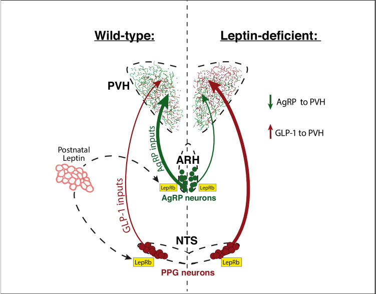
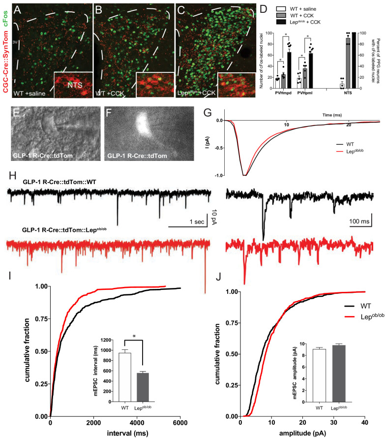
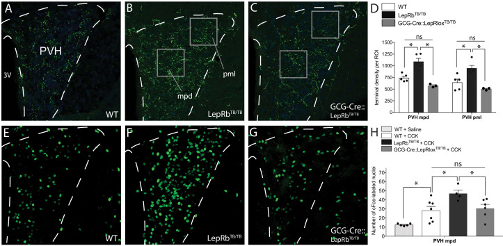
The nucleus of the solitary tract (NTS) is critical for the central integration of signals from visceral organs and contains preproglucagon (PPG) neurons, which express leptin receptors in the mouse and send direct projections to the paraventricular nucleus of the hypothalamus (PVH). Here, we visualized projections of PPG neurons in leptin-deficient mice and found that projections from PPG neurons are elevated compared with controls, and PPG projections were normalized by targeted rescue of leptin receptors in mice, which lack functional neuronal leptin receptors. Moreover, and mice displayed increased levels of neuronal activation in the PVH following vagal stimulation, and whole-cell patch recordings of GLP-1 receptor-expressing PVH neurons revealed enhanced excitatory neurotransmission, suggesting that leptin acts cell autonomously to suppress representation of excitatory afferents from PPG neurons, thereby diminishing the impact of visceral sensory information on GLP-1 receptor-expressing neurons in the PVH.

摘要

孤束核(NTS)对于内脏器官信号的中枢整合至关重要,其中包含前胰高血糖素(PPG)神经元,这些神经元在小鼠中表达瘦素受体,并向下丘脑室旁核(PVH)发出直接投射。在这里,我们可视化了瘦素缺乏小鼠中 PPG 神经元的投射,发现与对照组相比,PPG 神经元的投射增加,并且通过靶向拯救缺乏功能性神经元瘦素受体的小鼠中的瘦素受体,PPG 投射得到了正常化。此外,和 小鼠在迷走神经刺激后显示出 PVH 中神经元激活水平增加,并且表达 GLP-1 受体的 PVH 神经元的全细胞膜片钳记录显示兴奋性神经传递增强,表明瘦素自主作用以抑制来自 PPG 神经元的兴奋性传入的代表,从而减少内脏感觉信息对 PVH 中表达 GLP-1 受体的神经元的影响。

https://cdn.ncbi.nlm.nih.gov/pmc/blobs/04bb/7673779/2b7a0a42774e/elife-59857-fig8.jpg
https://cdn.ncbi.nlm.nih.gov/pmc/blobs/04bb/7673779/dae59c60573c/elife-59857-fig1.jpg
https://cdn.ncbi.nlm.nih.gov/pmc/blobs/04bb/7673779/5c81fe2210f4/elife-59857-fig2.jpg
https://cdn.ncbi.nlm.nih.gov/pmc/blobs/04bb/7673779/8b573a9ad644/elife-59857-fig2-figsupp1.jpg
https://cdn.ncbi.nlm.nih.gov/pmc/blobs/04bb/7673779/a635709b8983/elife-59857-fig3.jpg
https://cdn.ncbi.nlm.nih.gov/pmc/blobs/04bb/7673779/078b8d7e6d4b/elife-59857-fig4.jpg
https://cdn.ncbi.nlm.nih.gov/pmc/blobs/04bb/7673779/2340c0ac09c4/elife-59857-fig5.jpg
https://cdn.ncbi.nlm.nih.gov/pmc/blobs/04bb/7673779/f78908918393/elife-59857-fig6.jpg
https://cdn.ncbi.nlm.nih.gov/pmc/blobs/04bb/7673779/8a3b3a9eaaef/elife-59857-fig7.jpg
https://cdn.ncbi.nlm.nih.gov/pmc/blobs/04bb/7673779/2b7a0a42774e/elife-59857-fig8.jpg

相似文献

[1]
Leptin suppresses development of GLP-1 inputs to the paraventricular nucleus of the hypothalamus.

Elife. 2020-11-18

[2]
CCK stimulation of GLP-1 neurons involves α1-adrenoceptor-mediated increase in glutamatergic synaptic inputs.

Diabetes. 2011-9-1

[3]
Leptin directly depolarizes preproglucagon neurons in the nucleus tractus solitarius: electrical properties of glucagon-like Peptide 1 neurons.

Diabetes. 2010-6-3

[4]
Neurochemical characterization of body weight-regulating leptin receptor neurons in the nucleus of the solitary tract.

Endocrinology. 2012-8-6

[5]
Leptin receptor-expressing nucleus tractus solitarius neurons suppress food intake independently of GLP1 in mice.

JCI Insight. 2020-4-9

[6]
Fasting reduces the number of TRH immunoreactive neurons in the hypothalamic paraventricular nucleus of male rats, but not in mice.

Neurosci Lett. 2021-5-1

[7]
Divergent leptin signaling in proglucagon neurons of the nucleus of the solitary tract in mice and rats.

Endocrinology. 2008-2

[8]
Serotonergic modulation of the activity of GLP-1 producing neurons in the nucleus of the solitary tract in mouse.

Mol Metab. 2017-6-7

[9]
Upregulation of the brainstem preproglucagon system in the obese Zucker rat.

Brain Res. 2008-1-2

[10]
PPG neurons of the lower brain stem and their role in brain GLP-1 receptor activation.

Am J Physiol Regul Integr Comp Physiol. 2015-10-15

引用本文的文献

[1]
Vagal Oxytocin Receptors as Molecular Targets in Gut-Brain Signaling: Implications for Appetite, Satiety, Obesity, and Esophageal Motility-A Narrative Review.

Int J Mol Sci. 2025-8-13

[2]
Microglia are required for developmental specification of AgRP innervation in the hypothalamus of offspring exposed to maternal high-fat diet during lactation.

Elife. 2025-6-16

[3]
Postnatal hyperosmolality alters development of hypothalamic feeding circuits with context-specific changes in ingestive behavior.

iScience. 2025-3-24

[4]
Spotlight on the Mechanism of Action of Semaglutide.

Curr Issues Mol Biol. 2024-12-23

[5]
AgRP neurons mediate activity-dependent development of oxytocin connectivity and autonomic regulation.

Proc Natl Acad Sci U S A. 2024-12-3

[6]
History and future of leptin: Discovery, regulation and signaling.

Metabolism. 2024-12

[7]
Acute elevated dietary fat alone is not sufficient to decrease AgRP projections in the paraventricular nucleus of the hypothalamus in mice.

Sci Rep. 2024-8-29

[8]
Microglia are Required for Developmental Specification of AgRP Innervation in the Hypothalamus of Offspring Exposed to Maternal High-Fat Diet During Lactation.

bioRxiv. 2025-2-22

[9]
Obesity- and diet-induced plasticity in systems that control eating and energy balance.

Obesity (Silver Spring). 2024-8

[10]
Butterflies in the gut: the interplay between intestinal microbiota and stress.

J Biomed Sci. 2023-11-28

本文引用的文献

[1]
Preproglucagon Neurons in the Nucleus of the Solitary Tract Are the Main Source of Brain GLP-1, Mediate Stress-Induced Hypophagia, and Limit Unusually Large Intakes of Food.

Diabetes. 2018-10-2

[2]
GLP-1 neurons form a local synaptic circuit within the rodent nucleus of the solitary tract.

J Comp Neurol. 2018-9-19

[3]
Endogenous GLP-1 in lateral septum contributes to stress-induced hypophagia.

Physiol Behav. 2018-8-1

[4]
Enhanced AMPA Receptor Trafficking Mediates the Anorexigenic Effect of Endogenous Glucagon-like Peptide-1 in the Paraventricular Hypothalamus.

Neuron. 2017-11-15

[5]
Assessing the Functional Role of Leptin in Energy Homeostasis and the Stress Response in Vertebrates.

Front Endocrinol (Lausanne). 2017-4-7

[6]
Gut-Brain Cross-Talk in Metabolic Control.

Cell. 2017-2-23

[7]
Activation of murine pre-proglucagon-producing neurons reduces food intake and body weight.

J Clin Invest. 2017-3-1

[8]
Loss of MeCP2 disrupts cell autonomous and autocrine BDNF signaling in mouse glutamatergic neurons.

Elife. 2016-10-26

[9]
Sensory Neurons that Detect Stretch and Nutrients in the Digestive System.

Cell. 2016-6-30

[10]
GLP-1 and weight loss: unraveling the diverse neural circuitry.

Am J Physiol Regul Integr Comp Physiol. 2016-5-15

文献AI研究员

20分钟写一篇综述,助力文献阅读效率提升50倍

立即体验

用中文搜PubMed

大模型驱动的PubMed中文搜索引擎

马上搜索

推荐工具

医学文档翻译智能文献检索