• 文献检索
  • 文档翻译
  • 深度研究
  • 学术资讯
  • Suppr Zotero 插件Zotero 插件
  • 邀请有礼
  • 套餐&价格
  • 历史记录
应用&插件
Suppr Zotero 插件Zotero 插件浏览器插件Mac 客户端Windows 客户端微信小程序
定价
高级版会员购买积分包购买API积分包
服务
文献检索文档翻译深度研究API 文档MCP 服务
关于我们
关于 Suppr公司介绍联系我们用户协议隐私条款
关注我们

Suppr 超能文献

核心技术专利:CN118964589B侵权必究
粤ICP备2023148730 号-1Suppr @ 2026

文献检索

告别复杂PubMed语法,用中文像聊天一样搜索,搜遍4000万医学文献。AI智能推荐,让科研检索更轻松。

立即免费搜索

文件翻译

保留排版,准确专业,支持PDF/Word/PPT等文件格式,支持 12+语言互译。

免费翻译文档

深度研究

AI帮你快速写综述,25分钟生成高质量综述,智能提取关键信息,辅助科研写作。

立即免费体验

舌下腺导管的首次报告:动态磁共振涎腺造影成像及其临床应用

First Report of Sublingual Gland Ducts: Visualization by Dynamic MR Sialography and Its Clinical Application.

作者信息

Tanaka Tatsurou, Oda Masafumi, Wakasugi-Sato Nao, Joujima Takaaki, Miyamura Yuichi, Habu Manabu, Kodama Masaaki, Takahashi Osamu, Sago Teppei, Matsumoto-Takeda Shinobu, Nishida Ikuko, Tsurushima Hiroki, Otani Yasushi, Yoshiga Daigo, Sasaguri Masaaki, Morimoto Yasuhiro

机构信息

Division of Oral and Maxillofacial Radiology, Kyushu Dental University, Kitakyushu 803-8580, Japan.

Division of Maxillofacial Surgery, Kyushu Dental University, Kitakyushu 803-8580, Japan.

出版信息

J Clin Med. 2020 Nov 16;9(11):3676. doi: 10.3390/jcm9113676.

DOI:10.3390/jcm9113676
PMID:33207659
原文链接:https://pmc.ncbi.nlm.nih.gov/articles/PMC7698194/
Abstract

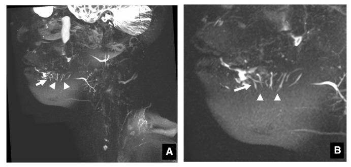
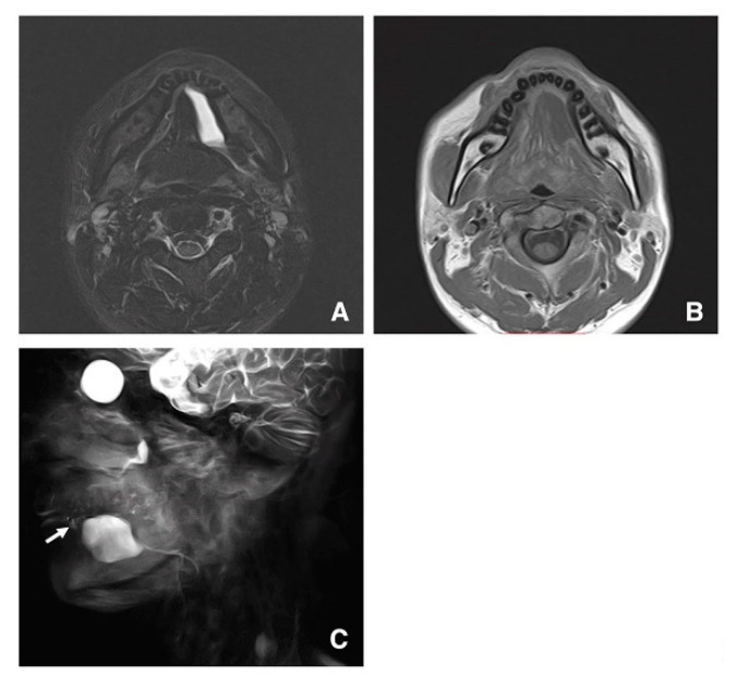
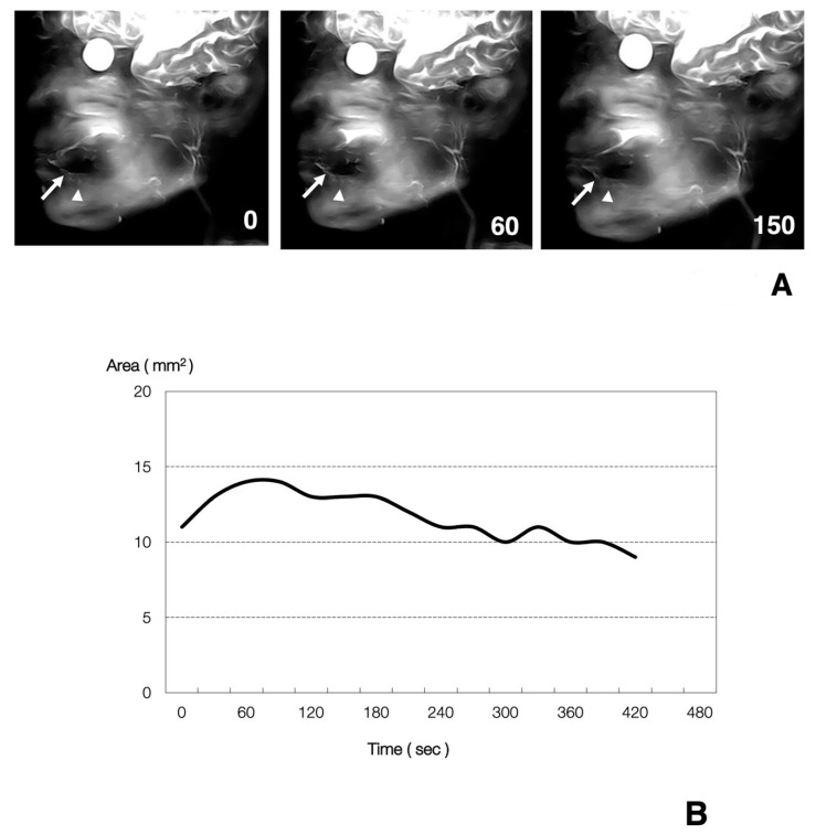
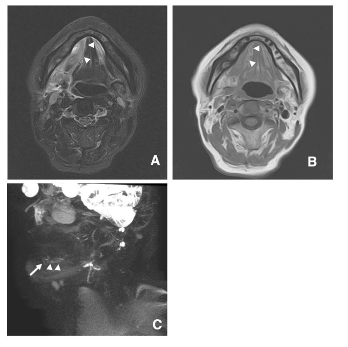
This study was done to determine whether the sublingual gland ducts could be visualized and/or their function assessed by MR sialography and dynamic MR sialography and to elucidate the clinical significance of the visualization and/or evaluation of the function of sublingual gland ducts by clinical application of these techniques. In 20 adult volunteers, 19 elderly volunteers, and 7 patients with sublingual gland disease, morphological and functional evaluations were done by MR sialography and dynamic MR sialography. Next, four parameters, including the time-dependent changes (change ratio) in the maximum area of the detectable sublingual gland ducts in dynamic MR sialographic images and data were analyzed. Sublingual gland ducts could be accurately visualized in 16 adult volunteers, 12 elderly volunteers, and 5 patients. No significant differences in the four parameters in detectable duct areas of sublingual glands were found among the three groups. In one patient with a ranula, the lesion could be correctly diagnosed as a ranula by MR sialography because the mass was clearly derived from sublingual gland ducts. This is the first report of successful visualization of sublingual gland ducts. In addition, the present study suggests that MR sialography can be more useful in the diagnosis of patients with lesions of sublingual gland ducts.

摘要

本研究旨在确定通过磁共振涎腺造影术和动态磁共振涎腺造影术能否观察舌下腺导管及其功能,并通过这些技术的临床应用阐明舌下腺导管可视化和/或功能评估的临床意义。对20名成年志愿者、19名老年志愿者和7名舌下腺疾病患者进行了磁共振涎腺造影术和动态磁共振涎腺造影术的形态学和功能评估。接下来,分析了四个参数,包括动态磁共振涎腺造影图像中可检测到的舌下腺导管最大面积的时间依赖性变化(变化率)和数据。在16名成年志愿者、12名老年志愿者和5名患者中,舌下腺导管能够被准确观察到。三组之间舌下腺可检测导管区域的四个参数没有显著差异。在一名患有舌下囊肿的患者中,磁共振涎腺造影术能够正确诊断该病变为舌下囊肿,因为肿块明显起源于舌下腺导管。这是首次成功观察到舌下腺导管的报告。此外,本研究表明磁共振涎腺造影术在舌下腺导管病变患者的诊断中可能更有用。

https://cdn.ncbi.nlm.nih.gov/pmc/blobs/16d6/7698194/6948ed3a54d2/jcm-09-03676-g004.jpg
https://cdn.ncbi.nlm.nih.gov/pmc/blobs/16d6/7698194/3b3a5a7258c3/jcm-09-03676-g001.jpg
https://cdn.ncbi.nlm.nih.gov/pmc/blobs/16d6/7698194/fcb157b19af4/jcm-09-03676-g002.jpg
https://cdn.ncbi.nlm.nih.gov/pmc/blobs/16d6/7698194/b9871e12881f/jcm-09-03676-g003.jpg
https://cdn.ncbi.nlm.nih.gov/pmc/blobs/16d6/7698194/6948ed3a54d2/jcm-09-03676-g004.jpg

相似文献

1
First Report of Sublingual Gland Ducts: Visualization by Dynamic MR Sialography and Its Clinical Application.舌下腺导管的首次报告:动态磁共振涎腺造影成像及其临床应用
J Clin Med. 2020 Nov 16;9(11):3676. doi: 10.3390/jcm9113676.
2
Comparison of the distribution of the Bartholin and/or Rivinus salivary ducts assessed with magnetic resonance-sialography in patients with ranula and in healthy subjects.用磁共振涎管造影评估的舌下腺和/或里维努斯唾液腺导管在舌下囊肿患者和健康受试者中的分布比较。
Quant Imaging Med Surg. 2024 Jan 3;14(1):397-407. doi: 10.21037/qims-23-948. Epub 2023 Nov 9.
3
Functional evaluations of the parotid and submandibular glands using dynamic magnetic resonance sialography.使用动态磁共振涎管造影术对腮腺和颌下腺进行功能评估。
Dentomaxillofac Radiol. 2007 May;36(4):218-23. doi: 10.1259/dmfr/27496576.
4
The functional evaluation of salivary glands using dynamic MR sialography following citric acid stimulation: a preliminary study.柠檬酸刺激后动态磁共振涎腺造影对涎腺的功能评估:一项初步研究。
Oral Surg Oral Med Oral Pathol Oral Radiol Endod. 2005 Sep;100(3):357-64. doi: 10.1016/j.tripleo.2004.11.053.
5
Virtual endoscopic view of salivary gland ducts using MR sialography data from three dimension fast asymmetric spin-echo (3D-FASE) sequences: a preliminary study.利用三维快速非对称自旋回波(3D-FASE)序列的磁共振涎管造影数据进行唾液腺导管的虚拟内镜观察:一项初步研究。
Oral Dis. 2002 Sep;8(5):268-74. doi: 10.1034/j.1601-0825.2002.01819.x.
6
3D MR sialography protocol for postradiotherapy follow-up of the salivary duct system.用于唾液腺导管系统放疗后随访的3D磁共振涎腺造影术方案。
J Magn Reson Imaging. 2006 Sep;24(3):556-62. doi: 10.1002/jmri.20659.
7
Dynamic magnetic resonance sialography for patients with xerostomia.口干症患者的动态磁共振涎管造影术
Oral Surg Oral Med Oral Pathol Oral Radiol Endod. 2008 Jul;106(1):115-23. doi: 10.1016/j.tripleo.2008.03.012. Epub 2008 May 27.
8
[MR sialography: evaluation of an ultra-fast sequence in consideration of a parallel acquisition technique and different functional conditions in patients with salivary gland diseases].[磁共振涎腺造影:考虑并行采集技术及唾液腺疾病患者不同功能状态下的超快速序列评估]
Rofo. 2007 Feb;179(2):153-8. doi: 10.1055/s-2006-927304.
9
Usefulness of Magnetic Resonance Sialography for the Evaluation of Radioactive Iodine-Induced Sialadenitis.磁共振涎管造影术在评估放射性碘诱发涎腺炎中的应用价值
Ann Surg Oncol. 2015 Dec;22 Suppl 3:S1007-13. doi: 10.1245/s10434-015-4681-2. Epub 2015 Aug 5.
10
MR sialography: the effect of a sialogogue and ductal occlusion in volunteers.磁共振涎管造影:催涎剂和导管阻塞对志愿者的影响。
Br J Radiol. 2008 Jul;81(967):583-6. doi: 10.1259/bjr/29344975. Epub 2008 Apr 28.

引用本文的文献

1
Comparison of the distribution of the Bartholin and/or Rivinus salivary ducts assessed with magnetic resonance-sialography in patients with ranula and in healthy subjects.用磁共振涎管造影评估的舌下腺和/或里维努斯唾液腺导管在舌下囊肿患者和健康受试者中的分布比较。
Quant Imaging Med Surg. 2024 Jan 3;14(1):397-407. doi: 10.21037/qims-23-948. Epub 2023 Nov 9.
2
A retrospective study of magnetic resonance sialography imaging applied in the prognostic evaluation of chronic obstructive parotitis.一项关于磁共振涎腺造影成像应用于慢性阻塞性腮腺炎预后评估的回顾性研究。
Quant Imaging Med Surg. 2023 Oct 1;13(10):6363-6373. doi: 10.21037/qims-22-1452. Epub 2023 Sep 12.
3

本文引用的文献

1
Significance of dynamic magnetic resonance sialography in prognostic evaluation of saline solution irrigation of the parotid gland for the treatment of xerostomia.动态磁共振涎腺造影在腮腺盐水灌注治疗口干症预后评估中的意义
J Oral Maxillofac Surg. 2010 Apr;68(4):768-76. doi: 10.1016/j.joms.2009.04.110. Epub 2009 Nov 13.
2
Magnetic resonance sialography using CISS and HASTE sequences in inflammatory salivary gland diseases: comparison with digital sialography.使用CISS和HASTE序列的磁共振涎管造影术在炎性唾液腺疾病中的应用:与数字涎管造影术的比较
Acta Radiol. 2010 Mar;51(2):156-63. doi: 10.3109/02841850903376306.
3
The role of magnetic resonance imaging and magnetic resonance sialography in the evaluation of salivary sialolithiasis: radiologic-endoscopic correlation.
磁共振成像和磁共振涎管造影在唾液腺涎石病评估中的作用:放射学与内镜的相关性
Pol J Radiol. 2022 Aug 5;87:e430-e437. doi: 10.5114/pjr.2022.118899. eCollection 2022.
4
Special Issue-"Diseases of the Salivary Glands".特刊 - “唾液腺疾病”。
J Clin Med. 2020 Nov 30;9(12):3886. doi: 10.3390/jcm9123886.
Autoimmune pancreatitis: imaging findings on contrast-enhanced MR, MRCP and dynamic secretin-enhanced MRCP.
自身免疫性胰腺炎:增强 MRI、MRCP 和动态促胰液素增强 MRCP 的影像学表现。
Radiol Med. 2009 Dec;114(8):1214-31. doi: 10.1007/s11547-009-0452-0. Epub 2009 Sep 30.
4
Preoperative evaluation of pancreatic cancer: comparison of gadolinium-enhanced dynamic MRI with MR cholangiopancreatography versus MDCT.胰腺癌的术前评估:钆增强动态磁共振成像联合磁共振胰胆管造影与多层螺旋CT的比较
J Magn Reson Imaging. 2009 Sep;30(3):586-95. doi: 10.1002/jmri.21889.
5
Radiation-induced xerostomia: objective evaluation of salivary gland injury using MR sialography.放射性口干症:使用磁共振涎管造影术对唾液腺损伤进行客观评估。
AJNR Am J Neuroradiol. 2009 Jan;30(1):53-8. doi: 10.3174/ajnr.A1322. Epub 2008 Oct 8.
6
Prospective evaluation of the diagnostic accuracy of secretin-enhanced magnetic resonance cholangiopancreaticography in suspected chronic pancreatitis.
Magn Reson Imaging. 2008 Dec;26(10):1367-73. doi: 10.1016/j.mri.2008.05.007. Epub 2008 Jun 25.
7
Dynamic magnetic resonance sialography for patients with xerostomia.口干症患者的动态磁共振涎管造影术
Oral Surg Oral Med Oral Pathol Oral Radiol Endod. 2008 Jul;106(1):115-23. doi: 10.1016/j.tripleo.2008.03.012. Epub 2008 May 27.
8
Diffusion-weighted magnetic resonance imaging to evaluate major salivary gland function before and after radiotherapy.采用扩散加权磁共振成像评估放疗前后大唾液腺功能。
Int J Radiat Oncol Biol Phys. 2008 Aug 1;71(5):1365-71. doi: 10.1016/j.ijrobp.2007.12.011. Epub 2008 Mar 20.
9
Functional evaluations of the parotid and submandibular glands using dynamic magnetic resonance sialography.使用动态磁共振涎管造影术对腮腺和颌下腺进行功能评估。
Dentomaxillofac Radiol. 2007 May;36(4):218-23. doi: 10.1259/dmfr/27496576.
10
Dynamic magnetic resonance sialography as a new diagnostic technique for patients with Sjögren's syndrome.动态磁共振涎腺造影术作为干燥综合征患者的一种新诊断技术。
Oral Dis. 2006 Jul;12(4):408-14. doi: 10.1111/j.1601-0825.2005.01215.x.