• 文献检索
  • 文档翻译
  • 深度研究
  • 学术资讯
  • Suppr Zotero 插件Zotero 插件
  • 邀请有礼
  • 套餐&价格
  • 历史记录
应用&插件
Suppr Zotero 插件Zotero 插件浏览器插件Mac 客户端Windows 客户端微信小程序
定价
高级版会员购买积分包购买API积分包
服务
文献检索文档翻译深度研究API 文档MCP 服务
关于我们
关于 Suppr公司介绍联系我们用户协议隐私条款
关注我们

Suppr 超能文献

核心技术专利:CN118964589B侵权必究
粤ICP备2023148730 号-1Suppr @ 2026

文献检索

告别复杂PubMed语法,用中文像聊天一样搜索,搜遍4000万医学文献。AI智能推荐,让科研检索更轻松。

立即免费搜索

文件翻译

保留排版,准确专业,支持PDF/Word/PPT等文件格式,支持 12+语言互译。

免费翻译文档

深度研究

AI帮你快速写综述,25分钟生成高质量综述,智能提取关键信息,辅助科研写作。

立即免费体验

胸部内镜超声检查(EBUS/EUS-b)在非小细胞肺癌患者纵隔分期中的临床应用:与PET/CT融合成像的比较——希腊的一项真实前瞻性研究

Clinical utility of thoracic endosonography (EBUS/EUS-b) in mediastinal staging of patients with non-small cell lung cancer: comparison with integrated PET/CT-a real-life prospective study in Greece.

作者信息

Chrysikos Serafeim, Gkiozos Ioannis, Dimakou Katerina, Zervas Eleftherios, Karampitsakos Theodoros, Anyfanti Maria, Tzouvelekis Argyrios, Samitas Konstantinos, Gaga Mina, Koulouris Nikolaos, Vasileiadis Ioannis, Syrigos Konstantinos

机构信息

5 Respiratory Medicine Department, "Sotiria" Chest Diseases Hospital, Athens, Greece.

3 Internal Medicine Department, "Sotiria" Chest Diseases Hospital, Medical School, National and Kapodistrian University of Athens, Athens, Greece.

出版信息

J Thorac Dis. 2020 Oct;12(10):5657-5666. doi: 10.21037/jtd-20-1735.

DOI:10.21037/jtd-20-1735
PMID:33209398
原文链接:https://pmc.ncbi.nlm.nih.gov/articles/PMC7656385/
Abstract

BACKGROUND

Accurate mediastinal staging in patients with non-small cell lung cancer (NSCLC) is crucial for the determination of optimal treatment management.

METHODS

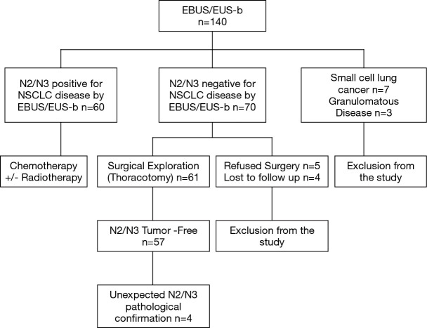
This was a real-life prospective study enrolling 140 patients between December 2016 and August 2018. We aimed to determine the clinical utility of EBUS/EUS-b in mediastinal staging of patients with NSCLC in comparison with integrated PET/CT. Furthermore, SUV cut-off value with the highest specificity/accuracy was evaluated. Subgroup analysis according to histological type was performed.

RESULTS

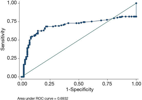
One hundred and thirty patients were eligible for analysis (mean age ± SD: 67.6±7.6, males 97). Three hundred different lymph node stations were sampled (272 through EBUS-TBNA and 28 through EUS-b FNA). Mean SUV of all malignant lymph nodes was 7.46 (SD =5.54). Sensitivity, specificity, PPV and NPV of EBUS/EUS-b for the identification of mediastinal malignant lymph nodes was 93.8%, 100%, 100%, and 93.4%, respectively. Accordingly, PET/CT yielded 92.2% sensitivity, 43.9% specificity, 64.8% PPV and 83.3% NPV. For adenocarcinoma (n=76) NPV were 86.2% with EBUS/EUS-b and 75% with PET/CT. NPV for squamous cell (n=46) was 100% with EBUS/EUS-b and 90.9% with PET/CT. EBUS/EUS-b staging yielded excellent agreement with final staging (97.5%, Tau 0.94, P<0.001). ROC curve analysis identified the value 4.95 as the optimal SUV cut-off value with the best specificity (87.4%) and accuracy (79%) (AUC 0.69; 95% CI: 0.73-0.84, P<0.001).

CONCLUSIONS

Thoracic endosonography is an excellent, minimally invasive tool yielding high sensitivity and diagnostic accuracy in mediastinal staging of patients with NSCLC. Implementation of both EBUS/EUS-b and PET/CT is necessary before any surgical intervention.

摘要

背景

准确的非小细胞肺癌(NSCLC)患者纵隔分期对于确定最佳治疗方案至关重要。

方法

这是一项在2016年12月至2018年8月期间纳入140例患者的真实前瞻性研究。我们旨在确定与PET/CT联合使用相比,EBUS/EUS-b在NSCLC患者纵隔分期中的临床应用价值。此外,还评估了具有最高特异性/准确性的SUV临界值。根据组织学类型进行亚组分析。

结果

130例患者符合分析条件(平均年龄±标准差:67.6±7.6岁,男性97例)。共采集了300个不同的淋巴结站(272个通过EBUS-TBNA,28个通过EUS-b FNA)。所有恶性淋巴结的平均SUV为7.46(标准差=5.54)。EBUS/EUS-b识别纵隔恶性淋巴结的敏感性、特异性、阳性预测值和阴性预测值分别为93.8%、100%、100%和93.4%。相应地,PET/CT的敏感性为92.2%,特异性为43.9%,阳性预测值为64.8%,阴性预测值为83.3%。对于腺癌(n=76),EBUS/EUS-b的阴性预测值为86.2%,PET/CT为75%。鳞状细胞癌(n=46)的EBUS/EUS-b阴性预测值为100%,PET/CT为90.9%。EBUS/EUS-b分期与最终分期具有极好的一致性(97.5%,Tau 0.94,P<0.001)。ROC曲线分析确定4.95为最佳SUV临界值,具有最佳特异性(87.4%)和准确性(79%)(AUC 0.69;95%CI:0.73-0.84,P<0.001)。

结论

胸部内镜超声是一种优秀的微创工具,在NSCLC患者纵隔分期中具有高敏感性和诊断准确性。在任何手术干预之前,同时实施EBUS/EUS-b和PET/CT是必要的。

https://cdn.ncbi.nlm.nih.gov/pmc/blobs/33cf/7656385/d26a9321b2d8/jtd-12-10-5657-f2.jpg
https://cdn.ncbi.nlm.nih.gov/pmc/blobs/33cf/7656385/426d3860ce49/jtd-12-10-5657-f1.jpg
https://cdn.ncbi.nlm.nih.gov/pmc/blobs/33cf/7656385/d26a9321b2d8/jtd-12-10-5657-f2.jpg
https://cdn.ncbi.nlm.nih.gov/pmc/blobs/33cf/7656385/426d3860ce49/jtd-12-10-5657-f1.jpg
https://cdn.ncbi.nlm.nih.gov/pmc/blobs/33cf/7656385/d26a9321b2d8/jtd-12-10-5657-f2.jpg

相似文献

1
Clinical utility of thoracic endosonography (EBUS/EUS-b) in mediastinal staging of patients with non-small cell lung cancer: comparison with integrated PET/CT-a real-life prospective study in Greece.胸部内镜超声检查(EBUS/EUS-b)在非小细胞肺癌患者纵隔分期中的临床应用:与PET/CT融合成像的比较——希腊的一项真实前瞻性研究
J Thorac Dis. 2020 Oct;12(10):5657-5666. doi: 10.21037/jtd-20-1735.
2
Combined endobronchial and esophageal endosonography for the diagnosis and staging of lung cancer: European Society of Gastrointestinal Endoscopy (ESGE) Guideline, in cooperation with the European Respiratory Society (ERS) and the European Society of Thoracic Surgeons (ESTS).支气管内与食管内超声联合用于肺癌诊断及分期:欧洲胃肠内镜学会(ESGE)指南,与欧洲呼吸学会(ERS)及欧洲胸外科医师学会(ESTS)合作制定
Endoscopy. 2015 Jun;47(6):545-59. doi: 10.1055/s-0034-1392040. Epub 2015 Jun 1.
3
Impact of F-FDG PET/CT, CT and EBUS/TBNA on preoperative mediastinal nodal staging of NSCLC.18F-氟脱氧葡萄糖正电子发射断层扫描/计算机断层扫描、计算机断层扫描和支气管内超声引导针吸活检术对非小细胞肺癌术前纵隔淋巴结分期的影响。
BMC Med Imaging. 2021 Mar 17;21(1):49. doi: 10.1186/s12880-021-00580-w.
4
Application of endobronchial ultrasound-guided transbronchial needle aspiration following integrated PET/CT in mediastinal staging of potentially operable non-small cell lung cancer.在综合PET/CT检查后,应用支气管内超声引导下经支气管针吸活检术对潜在可切除的非小细胞肺癌进行纵隔分期。
Chest. 2009 May;135(5):1280-1287. doi: 10.1378/chest.08-2019. Epub 2008 Dec 31.
5
Transbronchial and transesophageal fine-needle aspiration using an ultrasound bronchoscope in mediastinal staging of potentially operable lung cancer.超声支气管镜经支气管和经食管细针抽吸用于可手术肺癌的纵隔分期。
Chest. 2010 Oct;138(4):795-802. doi: 10.1378/chest.09-2100. Epub 2010 Mar 26.
6
Thoracic endosonography (EBUS/EUS-b) in the diagnosis of different intrathoracic diseases: A 4-year experience at a single-centre in Greece.胸腔内超声检查(EBUS/EUS-b)在不同胸腔内疾病诊断中的应用:希腊单中心 4 年经验。
Int J Clin Pract. 2021 Mar;75(3):e13684. doi: 10.1111/ijcp.13684. Epub 2020 Sep 29.
7
Minimally invasive endoscopic staging of suspected lung cancer.疑似肺癌的微创内镜分期
JAMA. 2008 Feb 6;299(5):540-6. doi: 10.1001/jama.299.5.540.
8
Excellence in non-small cell lung cancer staging by endobronchial-TBNA: Comparison with PET-CT and surgery.经支气管针吸活检术在非小细胞肺癌分期中的卓越表现:与PET-CT及手术的比较
Minim Invasive Ther Allied Technol. 2019 Aug;28(4):213-219. doi: 10.1080/13645706.2018.1513944. Epub 2018 Sep 27.
9
Is it necessary to sample the contralateral nodal stations by EBUS-TBNA in patients with lung cancer and clinical N0 / N1 on PET-CT?在 PET-CT 临床 N0/N1 肺癌患者中,是否有必要通过 EBUS-TBNA 对侧淋巴结站取样?
Lung Cancer. 2020 Apr;142:9-12. doi: 10.1016/j.lungcan.2020.01.014. Epub 2020 Jan 13.
10
Combined endobronchial and endoscopic ultrasound-guided fine needle aspiration for mediastinal nodal staging of lung cancer.经支气管内腔超声引导与内镜超声引导下细针抽吸术用于肺癌纵隔淋巴结分期。
Endoscopy. 2011 Dec;43(12):1082-9. doi: 10.1055/s-0030-1256766. Epub 2011 Oct 4.

引用本文的文献

1
Transesophageal endoscopic ultrasound with bronchoscope-guided fine-needle aspiration for diagnostic and staging purposes: a narrative review.经食管内镜超声检查联合支气管镜引导下细针穿刺活检用于诊断和分期:一项叙述性综述
J Thorac Dis. 2023 Sep 28;15(9):5088-5098. doi: 10.21037/jtd-23-681. Epub 2023 Aug 21.
2
Value of 18-fluorodeoxyglucose positron emission tomography/computed tomography (18F-FDG PET/CT) in the differential diagnosis of sarcoidosis and lung cancer with lymph node metastasis: a retrospective study.18-氟脱氧葡萄糖正电子发射断层扫描/计算机断层扫描(18F-FDG PET/CT)在结节病与肺癌伴淋巴结转移鉴别诊断中的价值:一项回顾性研究
Transl Lung Cancer Res. 2022 Sep;11(9):1926-1935. doi: 10.21037/tlcr-22-611.

本文引用的文献

1
The role of endobronchial ultrasound versus mediastinoscopy for non-small cell lung cancer.支气管内超声与纵隔镜检查在非小细胞肺癌诊断中的作用对比
J Thorac Dis. 2017 Mar;9(Suppl 2):S83-S97. doi: 10.21037/jtd.2017.03.102.
2
The Eighth Edition Lung Cancer Stage Classification.《第八版肺癌分期分类》
Chest. 2017 Jan;151(1):193-203. doi: 10.1016/j.chest.2016.10.010. Epub 2016 Oct 22.
3
Combined endobronchial and esophageal endosonography for the diagnosis and staging of lung cancer: European Society of Gastrointestinal Endoscopy (ESGE) Guideline, in cooperation with the European Respiratory Society (ERS) and the European Society of Thoracic Surgeons (ESTS).
支气管内与食管内超声联合用于肺癌诊断及分期:欧洲胃肠内镜学会(ESGE)指南,与欧洲呼吸学会(ERS)及欧洲胸外科医师学会(ESTS)合作制定
Endoscopy. 2015 Jun;47(6):545-59. doi: 10.1055/s-0034-1392040. Epub 2015 Jun 1.
4
Endoscopic ultrasound in the diagnosis and staging of lung cancer.内镜超声在肺癌的诊断和分期中的应用。
Endosc Ultrasound. 2014 Oct;3(4):205-12. doi: 10.4103/2303-9027.144510.
5
Combined endobronchial and transesophageal approach of an ultrasound bronchoscope for mediastinal staging of lung cancer.超声支气管镜联合经食管途径用于肺癌纵隔分期
PLoS One. 2014 Mar 14;9(3):e91893. doi: 10.1371/journal.pone.0091893. eCollection 2014.
6
Endosonographic mediastinal lymph node staging of lung cancer.肺癌的超声内镜纵隔淋巴结分期。
Chest. 2014 Aug;146(2):389-397. doi: 10.1378/chest.13-2349.
7
Revised ESTS guidelines for preoperative mediastinal lymph node staging for non-small-cell lung cancer.非小细胞肺癌术前纵隔淋巴结分期的修订版欧洲胸外科医师协会指南。
Eur J Cardiothorac Surg. 2014 May;45(5):787-98. doi: 10.1093/ejcts/ezu028. Epub 2014 Feb 26.
8
Early and locally advanced non-small-cell lung cancer (NSCLC): ESMO Clinical Practice Guidelines for diagnosis, treatment and follow-up.早期和局部晚期非小细胞肺癌(NSCLC):ESMO临床实践指南之诊断、治疗及随访
Ann Oncol. 2013 Oct;24 Suppl 6:vi89-98. doi: 10.1093/annonc/mdt241. Epub 2013 Jul 16.
9
Methods for staging non-small cell lung cancer: Diagnosis and management of lung cancer, 3rd ed: American College of Chest Physicians evidence-based clinical practice guidelines.非小细胞肺癌分期方法:肺癌的诊断与管理,第 3 版:美国胸科学会循证临床实践指南。
Chest. 2013 May;143(5 Suppl):e211S-e250S. doi: 10.1378/chest.12-2355.
10
The stage classification of lung cancer: Diagnosis and management of lung cancer, 3rd ed: American College of Chest Physicians evidence-based clinical practice guidelines.肺癌的分期:肺癌的诊断与管理,第 3 版:美国胸科医师学会循证临床实践指南。
Chest. 2013 May;143(5 Suppl):e191S-e210S. doi: 10.1378/chest.12-2354.