Tata Medical Centre, Kolkata, India.
Pacific Laboratories, Singapore, Singapore.
PLoS One. 2020 Nov 19;15(11):e0242058. doi: 10.1371/journal.pone.0242058. eCollection 2020.
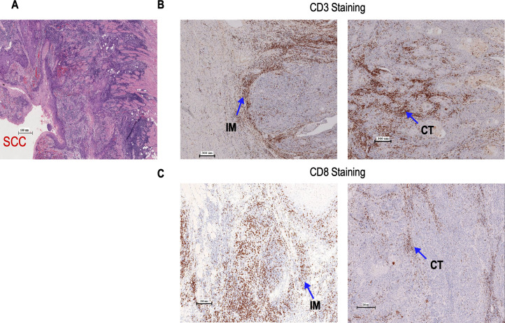
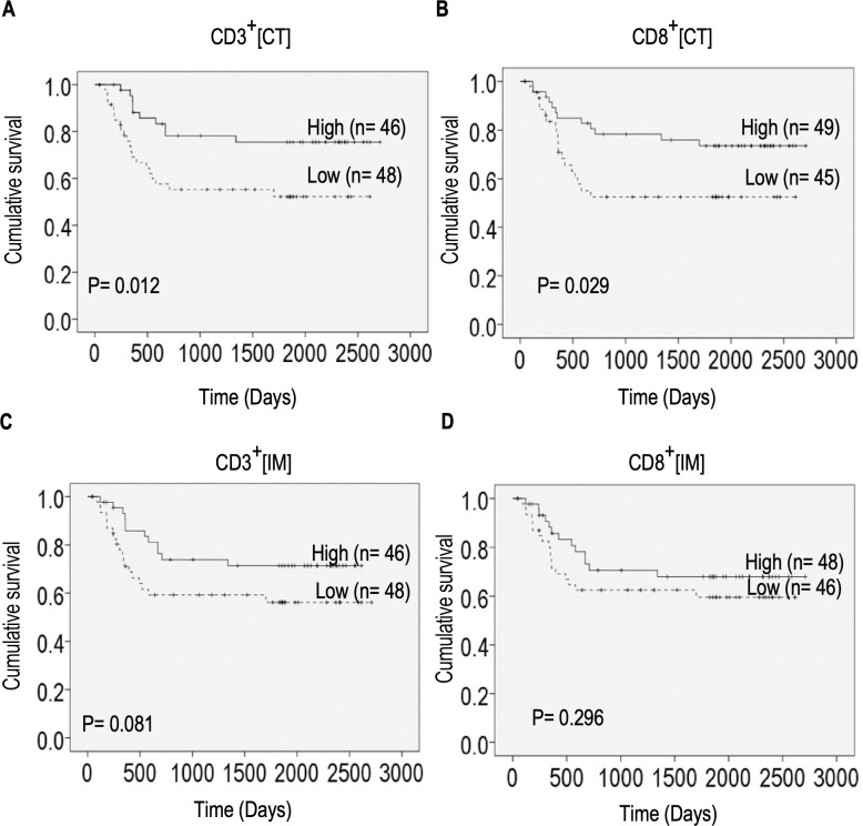
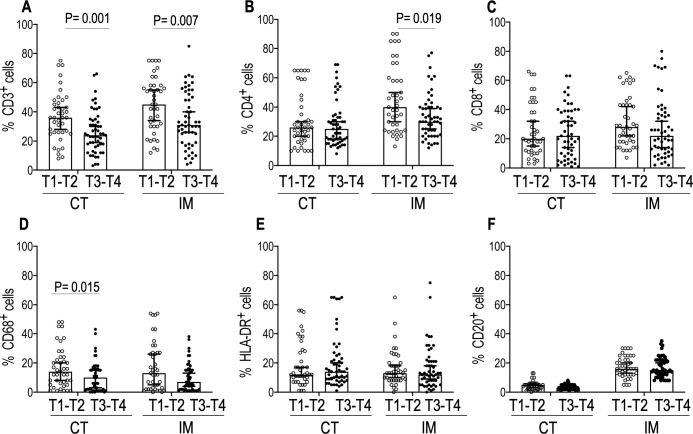
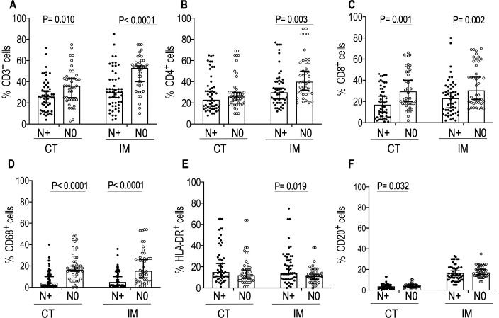
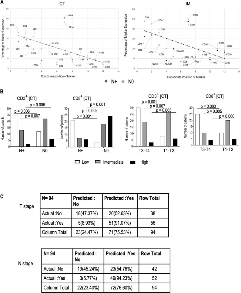
The tumor immune microenvironment is emerging as a critical player in predicting cancer prognosis and response to therapies. However, the prognostic value of tumor-infiltrating immune cells in Gingivo-Buccal Oral Squamous Cell Carcinoma (GBOSCC) and their association with tumor size or lymph node metastases status require further elucidation. To study the relationship of tumor-infiltrating immune cells with tumor size (T stage) and lymph node metastases (N stages), we analyzed the density of tumor-infiltrating immune cells in archived, whole tumor resections from 94 patients. We characterized these sections by immune-histochemistry using 12 markers and enumerated tumor-infiltrating immune cells at the invasive margins (IM) and centers of tumors (CT). We observed that a higher density of CD3+ cells in the IM and CT was associated with smaller tumor size (T1-T2 stage). Fewer CD3+ cells was associated with larger tumor size (T3-T4 stage). High infiltration of CD3+and CD8+ cells in IM and CT as well as high CD4+ cell infiltrates in the IM was significantly associated with the absence of lymph node metastases. High infiltrates of CD3+ and CD8+ cells in CT was associated with significantly improved survival. Our results illustrate that the densities and spatial distribution of CD3+ and CD8+ cell infiltrates in primary GBOSCC tumors is predictive of disease progression and survival. Based on our findings, we recommend incorporating immune cell quantification in the TNM classification and routine histopathology reporting of GBOSCC. Immune cell quantification in CT and IM may help predict the efficacy of future therapies.
肿瘤免疫微环境正成为预测癌症预后和对治疗反应的关键因素。然而,浸润肿瘤的免疫细胞在牙龈-颊口腔鳞状细胞癌(GBOSCC)中的预后价值及其与肿瘤大小或淋巴结转移状态的关系仍需进一步阐明。为了研究浸润肿瘤免疫细胞与肿瘤大小(T 分期)和淋巴结转移(N 分期)的关系,我们分析了 94 名患者存档的全肿瘤切除标本中浸润肿瘤免疫细胞的密度。我们使用 12 种标志物对这些切片进行免疫组织化学染色,并在肿瘤浸润边缘(IM)和中心(CT)计数浸润肿瘤的免疫细胞。我们观察到,IM 和 CT 中 CD3+细胞的密度越高,肿瘤体积越小(T1-T2 期)。CD3+细胞较少与较大的肿瘤大小(T3-T4 期)相关。IM 和 CT 中 CD3+和 CD8+细胞的高浸润以及 IM 中 CD4+细胞的高浸润与无淋巴结转移显著相关。CT 中 CD3+和 CD8+细胞的高浸润与显著改善的生存相关。我们的结果表明,原发性 GBOSCC 肿瘤中 CD3+和 CD8+细胞浸润的密度和空间分布可预测疾病进展和生存。基于我们的发现,我们建议在 GBOSCC 的 TNM 分类和常规组织病理学报告中纳入免疫细胞定量。CT 和 IM 中的免疫细胞定量可能有助于预测未来治疗的疗效。