Wagatsuma Kei, Sakata Muneyuki, Ishibashi Kenji, Hirayama Akira, Kawakami Hirofumi, Miwa Kenta, Suzuki Yukihisa, Ishii Kenji
Research Team for Neuroimaging, Tokyo Metropolitan Institute of Gerontology, 35-2 Sakae-cho, Itabashi-ku, Tokyo, 173-0015, Japan.
GE Healthcare Japan, 4-7-127 Asahigaoka, Hino, 191-8503, Japan.
EJNMMI Phys. 2020 Nov 23;7(1):70. doi: 10.1186/s40658-020-00337-4.
Silicon photomultiplier-positron emission tomography (SiPM-PET) has better sensitivity, spatial resolution, and timing resolution than photomultiplier tube (PMT)-PET. The present study aimed to clarify the advantages of SiPM-PET in F-fluoro-2-deoxy-D-glucose ([F]FDG) brain imaging in a head-to-head comparison with PMT-PET in phantom and clinical studies.
Contrast was calculated from images acquired from a Hoffman 3D brain phantom, and image noise and uniformity were calculated from images acquired from a pool phantom using SiPM- and PMT-PET. Sequential PMT-PET and SiPM-PET [F]FDG images were acquired over a period of 10 min from 22 controls and 10 patients. All images were separately normalized to a standard [F]FDG PET template, then the mean standardized uptake values (SUV) and Z-score were calculated using MIMneuro and CortexID Suite, respectively.
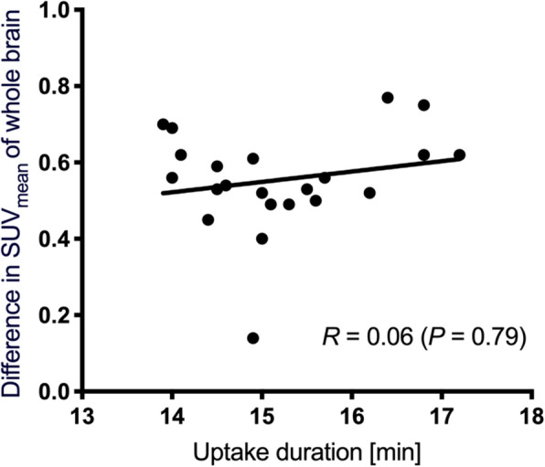
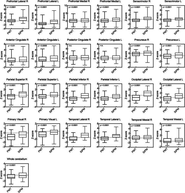
Image contrast, image noise, and uniformity in SiPM-PET changed 19.2, 3.5, and - 40.0% from PMT-PET, respectively. These physical indices of both PET scanners satisfied the criteria for acceptable image quality published by the Japanese Society of Nuclear Medicine of contrast > 55%, CV ≤ 15%, and SD ≤ 0.0249, respectively. Contrast was 70.0% for SiPM-PET without TOF and 59.5% for PMT-PET without TOF. The TOF improved contrast by 3.5% in SiPM-PET. The SUV using SiPM-PET was significantly higher than PMT-PET and did not correlate with a time delay. Z-scores were also significantly higher in images acquired from SiPM-PET (except for the bilateral posterior cingulate) than PMT-PET because the peak signal that was extracted by the calculation of Z-score in CortexID Suite was increased. The hypometabolic area in statistical maps was reduced and localized using SiPM-PET. The trend was independent of whether the images were derived from controls or patients.
The improved spatial resolution and sensitivity of SiPM-PET contributed to better image contrast and uniformity in brain [F]FDG images. The SiPM-PET offers better quality and more accurate quantitation of brain PET images. The SUV and Z-scores were higher in SiPM-PET than PMT-PET due to improved PVE. [F]FDG images acquired using SiPM-PET will help to improve diagnostic outcomes based on statistical image analysis because SiPM-PET would localize the distribution of glucose metabolism on Z-score maps.
硅光电倍增管正电子发射断层扫描(SiPM-PET)在灵敏度、空间分辨率和时间分辨率方面优于光电倍增管(PMT)-PET。本研究旨在通过在体模和临床研究中与PMT-PET进行直接比较,阐明SiPM-PET在F-氟-2-脱氧-D-葡萄糖([F]FDG)脑成像中的优势。
从霍夫曼3D脑体模获取的图像计算对比度,从水池体模获取的图像使用SiPM-PET和PMT-PET计算图像噪声和均匀性。在10分钟内从22名对照者和10名患者中依次采集PMT-PET和SiPM-PET [F]FDG图像。所有图像分别归一化为标准[F]FDG PET模板,然后分别使用MIMneuro和CortexID Suite计算平均标准化摄取值(SUV)和Z分数。
SiPM-PET的图像对比度、图像噪声和均匀性相对于PMT-PET分别变化了19.2%、3.5%和-40.0%。两种PET扫描仪的这些物理指标均符合日本核医学协会公布的可接受图像质量标准,对比度>55%,CV≤15%,SD≤0.0249。无飞行时间(TOF)的SiPM-PET对比度为70.0%,无TOF的PMT-PET对比度为59.5%。TOF使SiPM-PET的对比度提高了3.5%。使用SiPM-PET的SUV显著高于PMT-PET,且与时间延迟无关。在从SiPM-PET获取的图像(双侧后扣带回除外)中,Z分数也显著高于PMT-PET,因为在CortexID Suite中通过计算Z分数提取的峰值信号增加了。使用SiPM-PET可减少并定位统计图中的低代谢区域。该趋势与图像来源于对照者还是患者无关。
SiPM-PET空间分辨率和灵敏度的提高有助于改善脑[F]FDG图像的对比度和均匀性。SiPM-PET可提供更高质量和更准确的脑PET图像定量。由于部分容积效应(PVE)改善,SiPM-PET中的SUV和Z分数高于PMT-PET。使用SiPM-PET获取的[F]FDG图像将有助于基于统计图像分析改善诊断结果,因为SiPM-PET可在Z分数图上定位葡萄糖代谢分布。