• 文献检索
  • 文档翻译
  • 深度研究
  • 学术资讯
  • Suppr Zotero 插件Zotero 插件
  • 邀请有礼
  • 套餐&价格
  • 历史记录
应用&插件
Suppr Zotero 插件Zotero 插件浏览器插件Mac 客户端Windows 客户端微信小程序
定价
高级版会员购买积分包购买API积分包
服务
文献检索文档翻译深度研究API 文档MCP 服务
关于我们
关于 Suppr公司介绍联系我们用户协议隐私条款
关注我们

Suppr 超能文献

核心技术专利:CN118964589B侵权必究
粤ICP备2023148730 号-1Suppr @ 2026

文献检索

告别复杂PubMed语法,用中文像聊天一样搜索,搜遍4000万医学文献。AI智能推荐,让科研检索更轻松。

立即免费搜索

文件翻译

保留排版,准确专业,支持PDF/Word/PPT等文件格式,支持 12+语言互译。

免费翻译文档

深度研究

AI帮你快速写综述,25分钟生成高质量综述,智能提取关键信息,辅助科研写作。

立即免费体验

用于临床脑部研究的高分辨率硅光电倍增管飞行时间专用头部 PET 系统。

High-Resolution Silicon Photomultiplier Time-of-Flight Dedicated Head PET System for Clinical Brain Studies.

机构信息

Department of Radiology, Kindai University Faculty of Medicine, Osakasayama, Japan;

Division of Positron Emission Tomography, Institute of Advanced Clinical Medicine, Kindai University Hospital, Osakasayama, Japan; and.

出版信息

J Nucl Med. 2023 Jan;64(1):153-158. doi: 10.2967/jnumed.122.264080. Epub 2022 Jul 7.

DOI:10.2967/jnumed.122.264080
PMID:35798557
原文链接:https://pmc.ncbi.nlm.nih.gov/articles/PMC9841263/
Abstract

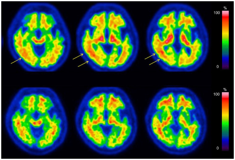
We acquired brain F-FDG and F-flutemetamol PET images using a time-of-flight system dedicated to the head (dhPET) and a conventional whole-body PET/CT (wbPET) system and evaluated the clinical superiority of dhPET over wbPET. There were 18 subjects for the F-FDG PET study and 17 subjects for the F-flutemetamol PET study. F-FDG PET images were first obtained using wbPET, followed by dhPET. F-flutemetamol PET images were first obtained using wbPET, followed by dhPET. Images acquired using dhPET and wbPET were compared by visual inspection, voxelwise analysis, and SUV ratio (SUVR). All F-FDG and F-flutemetamol images acquired using dhPET were judged as visually better than those acquired using wbPET. The voxelwise analysis demonstrated that accumulations in the cerebellum, in the lateral occipital cortices, and around the central sulcus area in dhPET F-FDG images were lower than those in wbPET F-FDG images, whereas accumulations around the ventricle systems were higher in dhPET F-FDG images than those in wbPET F-FDG images. Accumulations in the cerebellar dentate nucleus, in the midbrain, in the lateral occipital cortices, and around the central sulcus area in dhPET images were lower than those in wbPET images, whereas accumulations around the ventricle systems were higher in dhPET F-flutemetamol images than those in wbPET F-flutemetamol images. The mean cortical SUVRs of F-FDG and F-flutemetamol dhPET images were significantly higher than those of F-FDG and F-flutemetamol wbPET images, respectively. The dhPET images had better image quality by visual inspection and higher SUVRs than wbPET images. Although there were several regional accumulation differences between dhPET and wbPET images, understanding this phenomenon will enable full use of the features of this dhPET system in clinical practice.

摘要

我们使用专门用于头部的飞行时间系统(dhPET)和常规的全身 PET/CT(wbPET)系统获得脑 F-FDG 和 F-flutemetamol PET 图像,并评估了 dhPET 相对于 wbPET 的临床优势。有 18 名受试者进行 F-FDG PET 研究,17 名受试者进行 F-flutemetamol PET 研究。F-FDG PET 图像首先使用 wbPET 获得,然后使用 dhPET 获得。F-flutemetamol PET 图像首先使用 wbPET 获得,然后使用 dhPET 获得。通过视觉检查、体素分析和 SUV 比值(SUVr)比较 dhPET 和 wbPET 获得的图像。使用 dhPET 获得的所有 F-FDG 和 F-flutemetamol 图像均被判断为视觉效果优于使用 wbPET 获得的图像。体素分析表明,dhPET F-FDG 图像中小脑、外侧枕叶皮质和中央沟区域的蓄积低于 wbPET F-FDG 图像,而 dhPET F-FDG 图像中脑室系统周围的蓄积高于 wbPET F-FDG 图像。dhPET 图像中小脑齿状核、中脑、外侧枕叶皮质和中央沟区域的蓄积低于 wbPET 图像,而 dhPET F-flutemetamol 图像中脑室系统周围的蓄积高于 wbPET F-flutemetamol 图像。F-FDG 和 F-flutemetamol dhPET 图像的皮质 SUVr 平均值明显高于 F-FDG 和 F-flutemetamol wbPET 图像。dhPET 图像的视觉检查质量和 SUVr 均优于 wbPET 图像。尽管 dhPET 和 wbPET 图像之间存在一些区域蓄积差异,但了解这种现象将使我们能够在临床实践中充分利用这种 dhPET 系统的特点。

https://cdn.ncbi.nlm.nih.gov/pmc/blobs/3b31/9841263/04c976ebb1a2/jnumed.122.264080f6.jpg
https://cdn.ncbi.nlm.nih.gov/pmc/blobs/3b31/9841263/61a55d5d78bd/jnumed.122.264080absf1.jpg
https://cdn.ncbi.nlm.nih.gov/pmc/blobs/3b31/9841263/f890fe065e3b/jnumed.122.264080f1.jpg
https://cdn.ncbi.nlm.nih.gov/pmc/blobs/3b31/9841263/7a119636708f/jnumed.122.264080f2.jpg
https://cdn.ncbi.nlm.nih.gov/pmc/blobs/3b31/9841263/e6bedc448123/jnumed.122.264080f3.jpg
https://cdn.ncbi.nlm.nih.gov/pmc/blobs/3b31/9841263/47658c482b51/jnumed.122.264080f4.jpg
https://cdn.ncbi.nlm.nih.gov/pmc/blobs/3b31/9841263/8297d3cdd6f8/jnumed.122.264080f5.jpg
https://cdn.ncbi.nlm.nih.gov/pmc/blobs/3b31/9841263/04c976ebb1a2/jnumed.122.264080f6.jpg

相似文献

1
High-Resolution Silicon Photomultiplier Time-of-Flight Dedicated Head PET System for Clinical Brain Studies.用于临床脑部研究的高分辨率硅光电倍增管飞行时间专用头部 PET 系统。
J Nucl Med. 2023 Jan;64(1):153-158. doi: 10.2967/jnumed.122.264080. Epub 2022 Jul 7.
2
Visualization of small brain nuclei with a high-spatial resolution, clinically available whole-body PET scanner.利用高空间分辨率、临床可用的全身 PET 扫描仪对小脑中核进行可视化。
Ann Nucl Med. 2024 Feb;38(2):154-161. doi: 10.1007/s12149-023-01886-1. Epub 2023 Nov 21.
3
A cycle-consistent adversarial network for brain PET partial volume correction without prior anatomical information.一种无需先验解剖信息的基于循环一致性对抗网络的脑 PET 部分容积校正方法。
Eur J Nucl Med Mol Imaging. 2023 Jun;50(7):1881-1896. doi: 10.1007/s00259-023-06152-0. Epub 2023 Feb 20.
4
Optimization of image reconstruction conditions with phantoms for brain FDG and amyloid PET imaging.使用体模对脑部氟代脱氧葡萄糖(FDG)和淀粉样蛋白正电子发射断层扫描(PET)成像的图像重建条件进行优化。
Ann Nucl Med. 2016 Jan;30(1):18-28. doi: 10.1007/s12149-015-1024-0. Epub 2015 Sep 4.
5
Determination of optimal regularization factor in Bayesian penalized likelihood reconstruction of brain PET images using [ F]FDG and [ C]PiB.使用[F]FDG和[C]PiB对脑PET图像进行贝叶斯惩罚似然重建时最优正则化因子的确定
Med Phys. 2022 May;49(5):2995-3005. doi: 10.1002/mp.15593. Epub 2022 Mar 14.
6
Image reconstruction methods affect software-aided assessment of pathologies of [F]flutemetamol and [F]FDG brain-PET examinations in patients with neurodegenerative diseases.图像重建方法影响使用软件辅助评估神经退行性疾病患者 [F]氟替美莫和 [F]FDG 脑 PET 检查的病变情况。
Neuroimage Clin. 2020;28:102386. doi: 10.1016/j.nicl.2020.102386. Epub 2020 Aug 19.
7
The relationship between ring-type dedicated breast PET and immune microenvironment in early breast cancer.环形专用乳腺 PET 与早期乳腺癌免疫微环境的关系。
Breast Cancer Res Treat. 2019 Oct;177(3):651-657. doi: 10.1007/s10549-019-05339-0. Epub 2019 Jul 2.
8
Crossover evaluation of time-of-flight-based attenuation correction in brain F-FDG and F-flutemetamol PET.基于飞行时间的衰减校正技术在脑部F-FDG和F-氟代脱氧葡萄糖正电子发射断层显像中的交叉评估
Ann Nucl Med. 2025 Feb;39(2):189-198. doi: 10.1007/s12149-024-01986-6. Epub 2024 Sep 30.
9
Distribution pattern of FDG uptake using ring-type dedicated breast PET in comparison to whole-body PET/CT scanning in invasive breast cancer.环形专用乳腺 PET 与全身 PET/CT 扫描在浸润性乳腺癌中摄取 FDG 的分布模式比较。
Ann Nucl Med. 2019 Aug;33(8):570-578. doi: 10.1007/s12149-019-01364-7. Epub 2019 May 21.
10
Correlation of simultaneously acquired diffusion-weighted imaging and 2-deoxy-[18F] fluoro-2-D-glucose positron emission tomography of pulmonary lesions in a dedicated whole-body magnetic resonance/positron emission tomography system.在专用全身磁共振/正电子发射断层扫描系统中对肺部病变进行同时采集的弥散加权成像和 2-脱氧-[18F]氟-2-D-葡萄糖正电子发射断层扫描的相关性研究。
Invest Radiol. 2013 May;48(5):247-55. doi: 10.1097/RLI.0b013e31828d56a1.

引用本文的文献

1
Optimizing LM-DRAMA parameters and non-local means filtering to improve small-lesion detectability in SiPM-based TOF breast PET.优化LM-DRAMA参数和非局部均值滤波以提高基于硅光电倍增管的TOF乳腺PET中小病灶的可检测性。
Phys Eng Sci Med. 2025 Jul 22. doi: 10.1007/s13246-025-01598-y.
2
Recent Breakthroughs in PET-CT Multimodality Imaging: Innovations and Clinical Impact.PET-CT多模态成像的最新突破:创新与临床影响。
Bioengineering (Basel). 2024 Nov 30;11(12):1213. doi: 10.3390/bioengineering11121213.
3
Crossover evaluation of time-of-flight-based attenuation correction in brain F-FDG and F-flutemetamol PET.

本文引用的文献

1
Meaningful benefits: a framework to assess disease-modifying therapies in preclinical and early Alzheimer's disease.有意义的获益:评估早期阿尔茨海默病中疾病修饰疗法的框架。
Alzheimers Res Ther. 2022 Apr 19;14(1):54. doi: 10.1186/s13195-022-00984-y.
2
Initial evaluation of a new maximum-likelihood attenuation correction factor-based attenuation correction for time-of-flight brain PET.基于最大似然衰减校正因子的飞行时间脑 PET 新衰减校正的初步评估。
Ann Nucl Med. 2022 Apr;36(4):420-426. doi: 10.1007/s12149-022-01721-z. Epub 2022 Feb 9.
3
Relationship between F-18 florbetapir uptake in occipital lobe and neurocognitive performance in Alzheimer's disease.
基于飞行时间的衰减校正技术在脑部F-FDG和F-氟代脱氧葡萄糖正电子发射断层显像中的交叉评估
Ann Nucl Med. 2025 Feb;39(2):189-198. doi: 10.1007/s12149-024-01986-6. Epub 2024 Sep 30.
4
Can the Newer Model of Breast-Specific Positron Emission Tomography Reduce the "Blind Area"?新型乳腺专用正电子发射断层扫描模型能否减少“盲区”?
Diagnostics (Basel). 2024 Sep 19;14(18):2068. doi: 10.3390/diagnostics14182068.
5
Generating PET Attenuation Maps via Sim2Real Deep Learning-Based Tissue Composition Estimation Combined with MLACF.通过基于 Sim2Real 深度学习的组织成分估计与 MLACF 相结合生成 PET 衰减图。
J Imaging Inform Med. 2024 Feb;37(1):167-179. doi: 10.1007/s10278-023-00902-0. Epub 2024 Jan 10.
6
Visualization of small brain nuclei with a high-spatial resolution, clinically available whole-body PET scanner.利用高空间分辨率、临床可用的全身 PET 扫描仪对小脑中核进行可视化。
Ann Nucl Med. 2024 Feb;38(2):154-161. doi: 10.1007/s12149-023-01886-1. Epub 2023 Nov 21.
7
The quest for multifunctional and dedicated PET instrumentation with irregular geometries.追求具有不规则几何形状的多功能、专用 PET 仪器。
Ann Nucl Med. 2024 Jan;38(1):31-70. doi: 10.1007/s12149-023-01881-6. Epub 2023 Nov 12.
8
Organ-Specific Positron Emission Tomography Scanners for Breast Imaging: Comparison between the Performances of Prior and Novel Models.用于乳腺成像的器官特异性正电子发射断层扫描仪:既往型号与新型号性能比较
Diagnostics (Basel). 2023 Mar 13;13(6):1079. doi: 10.3390/diagnostics13061079.
9
Attenuation correction for phantom tests: an alternative to maximum-likelihood attenuation correction factor-based correction for clinical studies in time-of-flight PET.衰减校正的体模测试:替代最大似然衰减校正因子的校正方法,用于飞行时间 PET 临床研究。
Ann Nucl Med. 2022 Nov;36(11):998-1006. doi: 10.1007/s12149-022-01788-8. Epub 2022 Sep 28.
阿尔茨海默病患者枕叶 F-18 氟比他滨摄取与神经认知表现的关系。
Jpn J Radiol. 2021 Oct;39(10):984-993. doi: 10.1007/s11604-021-01132-6. Epub 2021 May 21.
4
PET Imaging in Neurodegeneration and Neuro-oncology: Variants and Pitfalls.正电子发射断层扫描成像在神经退行性疾病和神经肿瘤学中的应用:变异与陷阱。
Semin Nucl Med. 2021 Sep;51(5):408-418. doi: 10.1053/j.semnuclmed.2021.03.003. Epub 2021 Apr 3.
5
Alzheimer's disease.阿尔茨海默病。
Lancet. 2021 Apr 24;397(10284):1577-1590. doi: 10.1016/S0140-6736(20)32205-4. Epub 2021 Mar 2.
6
Brain [F-18]FDG PET for Clinical Dementia Workup: Differential Diagnosis of Alzheimer's Disease and Other Types of Dementing Disorders.用于临床痴呆症检查的脑[F-18]氟代脱氧葡萄糖正电子发射断层显像:阿尔茨海默病与其他类型痴呆症的鉴别诊断
Semin Nucl Med. 2021 May;51(3):230-240. doi: 10.1053/j.semnuclmed.2021.01.002. Epub 2021 Feb 2.
7
Quantitative Evaluation of F-Flutemetamol PET in Patients With Cognitive Impairment and Suspected Alzheimer's Disease: A Multicenter Study.认知障碍和疑似阿尔茨海默病患者中F-氟代去甲肾上腺素PET的定量评估:一项多中心研究
Front Neurol. 2021 Jan 13;11:578753. doi: 10.3389/fneur.2020.578753. eCollection 2020.
8
Future Imaging in Dementia.痴呆症的未来影像学
Semin Nucl Med. 2021 May;51(3):303-308. doi: 10.1053/j.semnuclmed.2020.12.001. Epub 2020 Dec 20.
9
Direct comparison of brain [F]FDG images acquired by SiPM-based and PMT-based PET/CT: phantom and clinical studies.基于硅光电倍增管(SiPM)和基于光电倍增管(PMT)的PET/CT所采集的脑部[F]FDG图像的直接比较:体模和临床研究。
EJNMMI Phys. 2020 Nov 23;7(1):70. doi: 10.1186/s40658-020-00337-4.
10
Regional gray matter-dedicated SUVR with 3D-MRI detects positive amyloid deposits in equivocal amyloid PET images.采用 3D-MRI 技术的区域性灰质特异性 SUVR 可探测到疑似淀粉样 PET 图像中的阳性淀粉样沉积物。
Ann Nucl Med. 2020 Nov;34(11):856-863. doi: 10.1007/s12149-020-01513-3. Epub 2020 Aug 27.