Capra Davide, Monti Caterina Beatrice, Luporini Alberto Gianluigi, Lombardi Fabrizio, Gumina Calogero, Sironi Andrea, Asti Emanuele Luigi Giuseppe, Bonavina Luigi, Secchi Francesco, Sardanelli Francesco
Department of Biomedical Sciences for Health, Università Degli Studi Di Milano, Via Mangiagalli 31, 20133, Milano, Italy.
Unit of Medical Oncology, IRCCS Policlinico San Donato, Via Morandi 30, 20097, San Donato Milanese, Italy.
Insights Imaging. 2020 Nov 23;11(1):120. doi: 10.1186/s13244-020-00922-2.
We aimed to assess extracellular volume (ECV) through non-gated, contrast-enhanced computed tomography (CT) before and after radiation therapy (RT) in patients with esophageal cancer (EC).
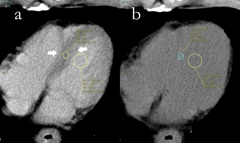
EC patients who had undergone CT before and after RT were retrospectively assessed. Patients with preexisting cardiovascular disease or with heavily artifacted CT were excluded. ECV was calculated using density values for the myocardial septum and blood pool. Data were reported as mean and standard deviation or median and interquartile range according to their distribution; t test or Wilcoxon and Pearson r or Spearman ρ were subsequently used.
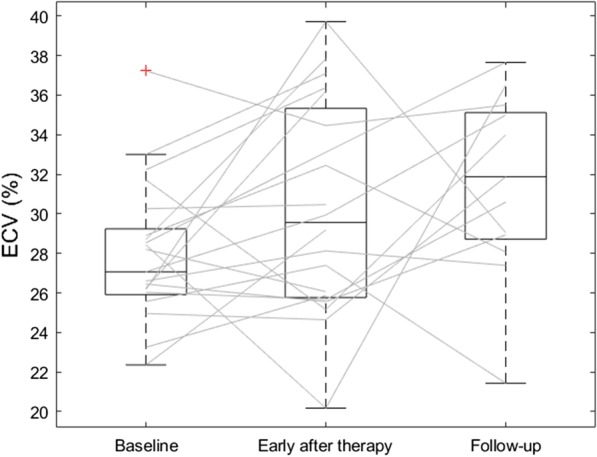
Twenty-one patients with stage ≥ IB EC, aged 64 ± 18 years, were included. Mean and maximum RT doses were 21.2 Gy (16.9-24.1) and 42.5 Gy (41.8-49.2), respectively. At baseline (n = 21), hematocrit was 39% ± 4%, ECV 27.9% ± 3.5%; 35 days (30-38) after RT (n = 20), hematocrit was 36% ± 4%, lower than at baseline (p = 0.002), ECV 30.3% ± 8.3%, higher than at baseline (p = 0.081); at follow-up 420 days (244-624) after RT (n = 13), hematocrit was 36% ± 5%, lower than at baseline (p = 0.030), ECV 31.4% ± 4.5%, higher than at baseline (p = 0.011). No patients showed signs of overt cardiotoxicity. ECV early after RT was moderately positively correlated with maximum RT dose (ρ = 0.50, p = 0.036).
In EC patients, CT-derived myocardial ECV was increased after RT and may thus appear as a potential early biomarker of cardiotoxicity.
我们旨在通过非门控对比增强计算机断层扫描(CT)评估食管癌(EC)患者放疗(RT)前后的细胞外液体积(ECV)。
对接受放疗前后CT检查的EC患者进行回顾性评估。排除既往有心血管疾病或CT图像严重伪影的患者。使用心肌间隔和血池的密度值计算ECV。数据根据其分布以均值和标准差或中位数和四分位数间距报告;随后使用t检验或Wilcoxon检验以及Pearson r或Spearman ρ检验。
纳入21例IB期及以上EC患者,年龄64±18岁。平均和最大放疗剂量分别为21.2 Gy(16.9 - 24.1)和42.5 Gy(41.8 - 49.2)。基线时(n = 21),血细胞比容为39%±4%,ECV为27.9%±3.5%;放疗后35天(30 - 38)(n = 20),血细胞比容为36%±4%,低于基线(p = 0.002),ECV为30.3%±8.3%,高于基线(p = 0.081);放疗后420天(244 - 624)随访时(n = 13),血细胞比容为36%±5%,低于基线(p = 0.030),ECV为31.4%±4.5%,高于基线(p = 0.011)。无患者出现明显心脏毒性迹象。放疗后早期ECV与最大放疗剂量呈中度正相关(ρ = 0.50,p = 0.036)。
在EC患者中,放疗后CT衍生的心肌ECV增加,因此可能是心脏毒性的潜在早期生物标志物。