Department of Ophthalmology and Visual Sciences, University of Utah, Salt Lake City, UT, USA.
Department of Bioengineering, University of Utah, Salt Lake City, UT, USA.
J Physiol. 2021 Jan;599(2):571-592. doi: 10.1113/JP281011. Epub 2020 Dec 12.
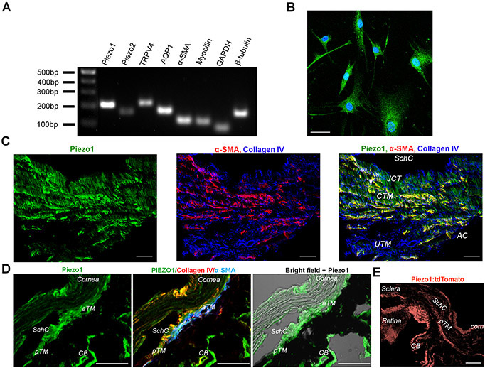
Trabecular meshwork (TM) is a highly mechanosensitive tissue in the eye that regulates intraocular pressure through the control of aqueous humour drainage. Its dysfunction underlies the progression of glaucoma but neither the mechanisms through which TM cells sense pressure nor their role in aqueous humour outflow are understood at the molecular level. We identified the Piezo1 channel as a key TM transducer of tensile stretch, shear flow and pressure. Its activation resulted in intracellular signals that altered organization of the cytoskeleton and cell-extracellular matrix contacts and modulated the trabecular component of aqueous outflow whereas another channel, TRPV4, mediated a delayed mechanoresponse. This study helps elucidate basic mechanotransduction properties that may contribute to intraocular pressure regulation in the vertebrate eye.
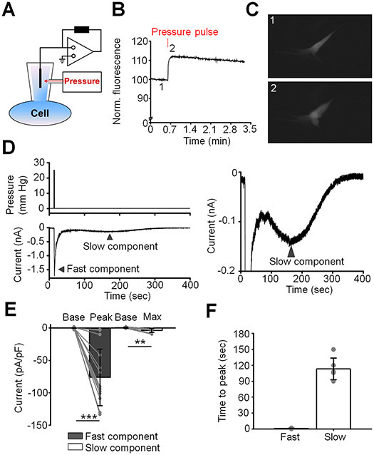
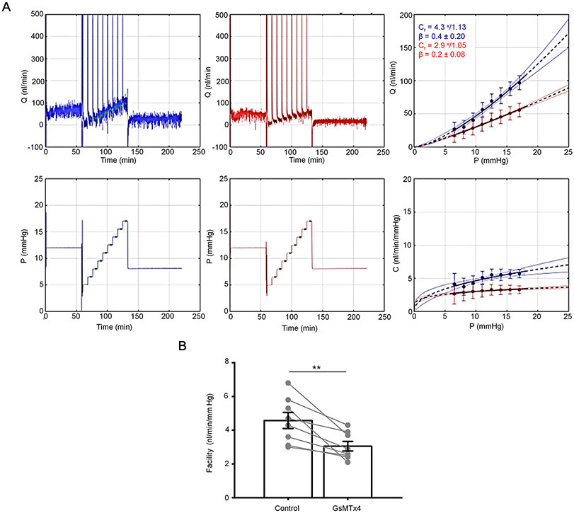
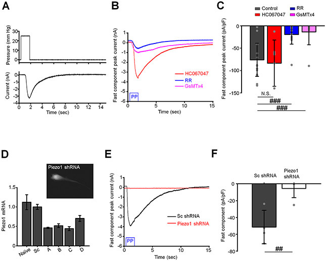
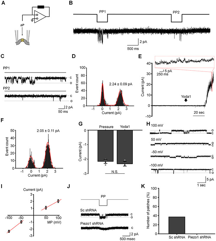
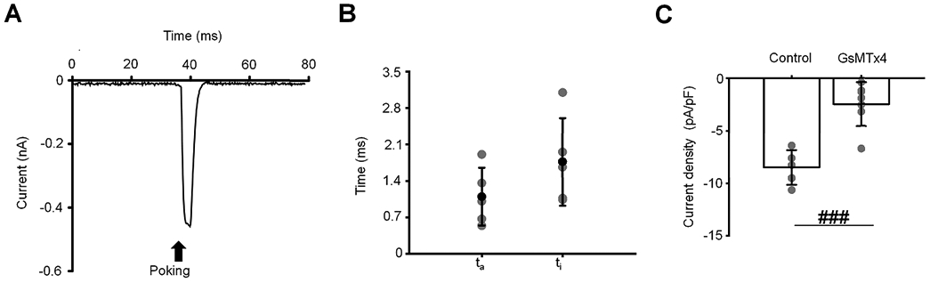
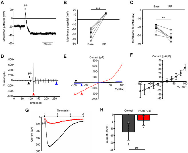
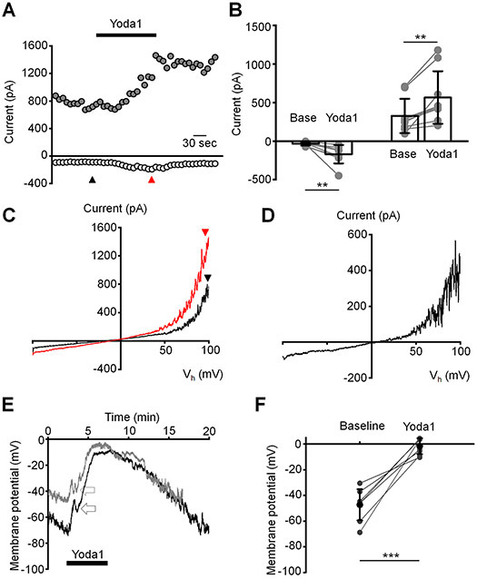
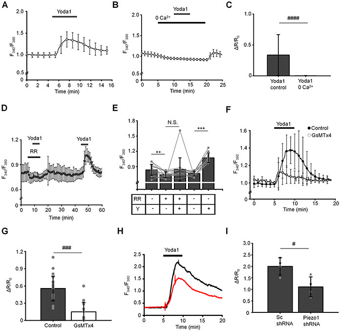
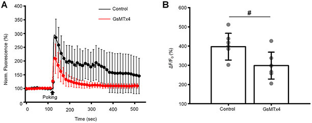
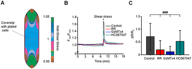
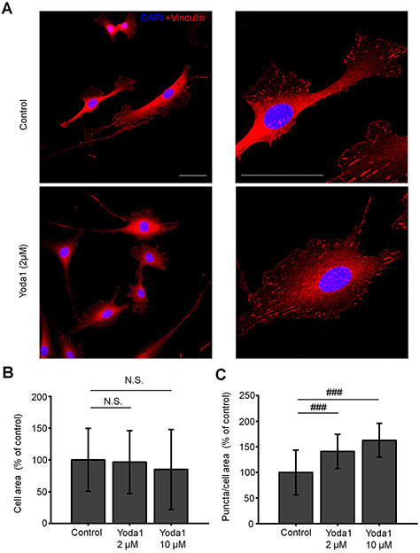
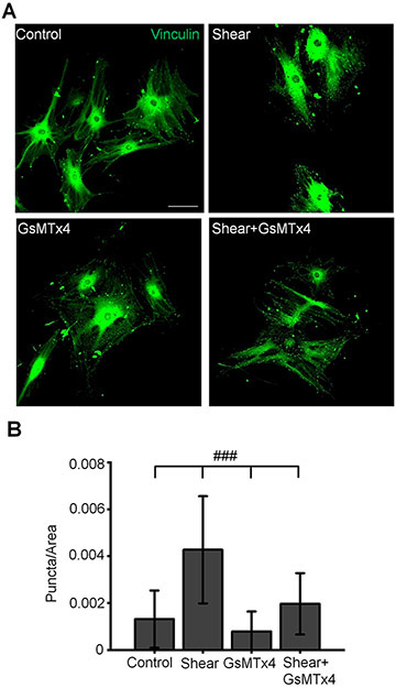
Chronic elevations in intraocular pressure (IOP) can cause blindness by compromising the function of trabecular meshwork (TM) cells in the anterior eye, but how these cells sense and transduce pressure stimuli is poorly understood. Here, we demonstrate functional expression of two mechanically activated channels in human TM cells. Pressure-induced cell stretch evoked a rapid increase in transmembrane current that was inhibited by antagonists of the mechanogated channel Piezo1, Ruthenium Red and GsMTx4, and attenuated in Piezo1-deficient cells. The majority of TM cells exhibited a delayed stretch-activated current that was mediated independently of Piezo1 by TRPV4 (transient receptor potential cation channel, subfamily V, member 4) channels. Piezo1 functions as the principal TM transducer of physiological levels of shear stress, with both shear and the Piezo1 agonist Yoda1 increasing the number of focal cell-matrix contacts. Analysis of TM-dependent fluid drainage from the anterior eye showed significant inhibition by GsMTx4. Collectively, these results suggest that TM mechanosensitivity utilizes kinetically, regulatory and functionally distinct pressure transducers to inform the cells about force-sensing contexts. Piezo1-dependent control of shear flow sensing, calcium homeostasis, cytoskeletal dynamics and pressure-dependent outflow suggests potential for a novel therapeutic target in treating glaucoma.
小梁网(TM)是眼睛中一种高度机械敏感的组织,通过控制房水引流来调节眼内压。其功能障碍是青光眼进展的基础,但 TM 细胞感知压力的机制及其在房水流出中的作用在分子水平上尚不清楚。我们发现 Piezo1 通道是 TM 细胞拉伸应变、切变流和压力的关键转导器。其激活导致细胞内信号改变细胞骨架和细胞-细胞外基质接触的组织,并调节房水流出的小梁成分,而另一种通道 TRPV4 介导延迟的机械反应。这项研究有助于阐明基本的机械转导特性,这些特性可能有助于脊椎动物眼睛的眼压调节。
眼内压(IOP)的慢性升高会通过损害前眼部小梁网(TM)细胞的功能导致失明,但这些细胞如何感知和转导压力刺激知之甚少。在这里,我们证明了两种机械激活通道在人 TM 细胞中的功能表达。压力诱导的细胞拉伸引起跨膜电流的快速增加,该电流被机械门控通道 Piezo1 的拮抗剂 Ruthenium Red 和 GsMTx4 抑制,并在 Piezo1 缺陷细胞中减弱。大多数 TM 细胞表现出延迟的拉伸激活电流,该电流独立于 Piezo1 由 TRPV4(瞬时受体电位阳离子通道,亚家族 V,成员 4)通道介导。Piezo1 是生理水平剪切应力的主要 TM 转导器,剪切应力和 Piezo1 激动剂 Yoda1 都增加了焦点细胞-基质接触的数量。分析前眼部依赖 TM 的液体引流显示 GsMTx4 有显著抑制作用。总之,这些结果表明,TM 的机械敏感性利用动力学上、调节上和功能上不同的压力传感器来告知细胞有关力感应的情况。Piezo1 依赖的剪切流感应、钙稳态、细胞骨架动力学和压力依赖性流出的控制表明,在治疗青光眼方面具有潜在的新治疗靶点。