Experimental Pathology Laboratory, Laboratory of Pathology, Center for Cancer Research, National Cancer Institute, National Institutes of Health, MSC1500, Bethesda, MD, 20892, USA.
Department of Pathology, Keimyung University Scholl of Medicine and Institute for Cancer Research, Dongsan Medical Center, Daegu, 42601, Republic of Korea.
J Transl Med. 2020 Nov 23;18(1):443. doi: 10.1186/s12967-020-02618-z.
The tumor microenvironment (TME) is a critical player in tumor progression, metastasis and therapy outcomes. Tumor-associated macrophages (TAMs) are a well-recognized core element of the TME and generally characterized as M2-like macrophages. TAMs are believed to contribute to tumor progression, but the mechanism behind this remains unclear. We aimed to investigate the clinical, angiogenic, and lymphangiogenic significance of TAMs in non-small cell lung cancer (NSCLC).
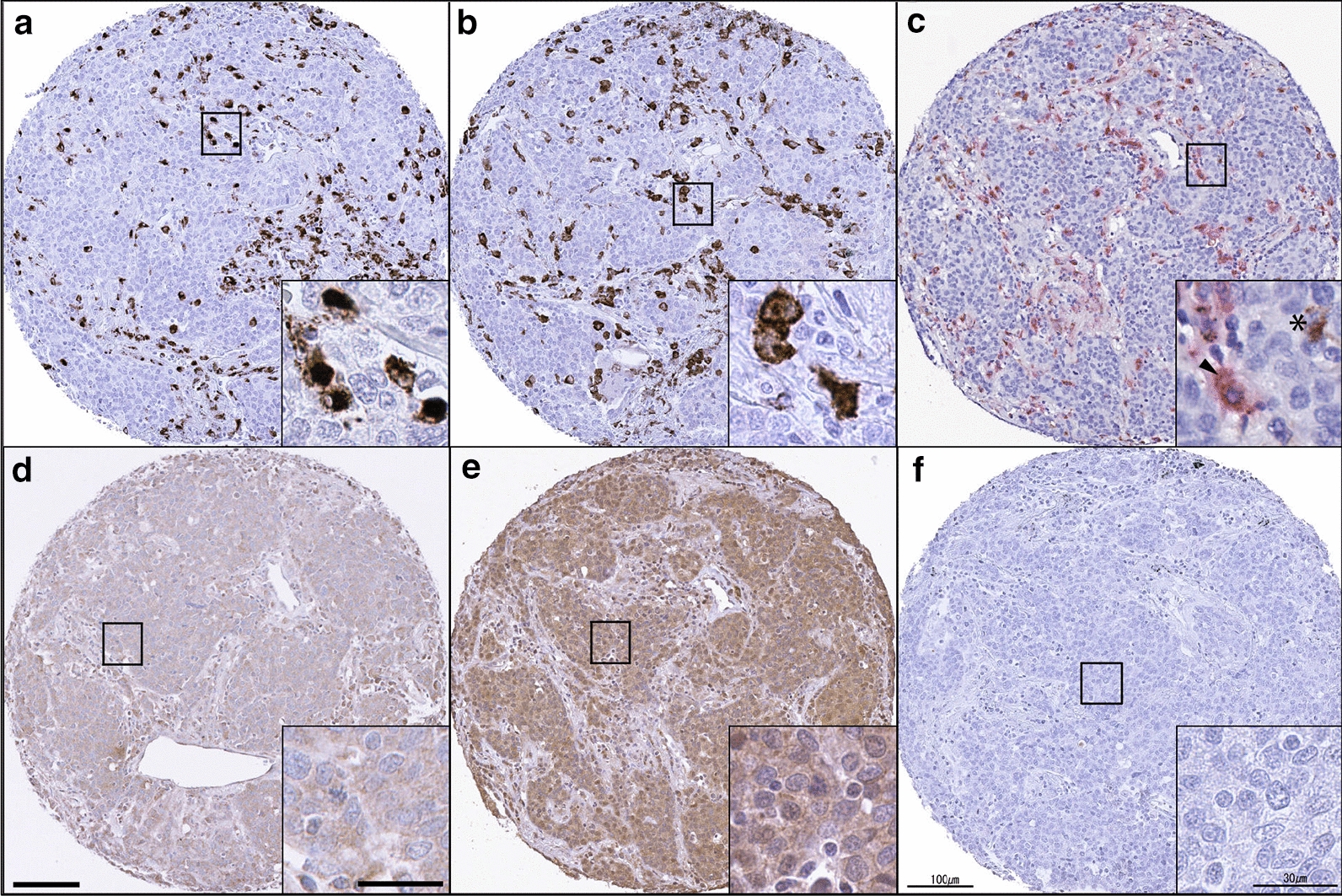
Utilizing combined immunohistochemistry and digital image analysis, we assessed CD68, CD163, VEGF-A, and VEGF-C expression in 349 patients with NSCLC. Subsequently, the potential association between M2 TAMs and angiogenic VEGF-A and/or lymphangiogenic VEGF-C was evaluated for its prognostic value. Furthermore, the effects of M2 TAMs on angiogenesis and lymphangiogenesis were explored via an in vitro co-culture system.
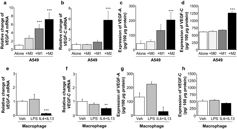
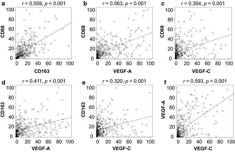
CD68 and CD163 expression were found to directly correlate with VEGF-A and/or VEGF-C expression (all p < 0.001). Furthermore, elevated M2 ratio (CD163+/CD68+) was significantly associated with poor overall survival (p = 0.023). Dual expression of M2 ratio and VEGF-C (M2 ratioVEGF-C) was correlated with worse overall survival (p = 0.033). Multivariate analysis revealed that M2 ratio [HR (95% CI) = 1.53 (1.01-2.33), p = 0.046] and combined M2 ratioVEGF-C expression [HR (95% CI) = 2.01 (1.28-3.16), p = 0.003] were independent predictors of poor overall survival. Notably, we confirmed that M2 macrophages significantly enhanced the protein and mRNA expression of both VEGF-A and VEGF-C, while M1 macrophages induced only mRNA expression of VEGF-A in A549 cells.
This study suggests that TAMs are significantly associated with angiogenesis and lymphangiogenesis, contributing to the progression of NSCLC. Furthermore, elevated M2 ratio, similar to combined high M2 ratio and high VEGF-C expression, is a strong indicator of poor prognosis in patients with NSCLC, providing insight for future TAM-based immunotherapy strategies.
肿瘤微环境(TME)是肿瘤进展、转移和治疗结果的关键因素。肿瘤相关巨噬细胞(TAMs)是 TME 的一个公认的核心组成部分,通常被特征化为 M2 样巨噬细胞。TAMs 被认为有助于肿瘤进展,但这背后的机制尚不清楚。我们旨在研究非小细胞肺癌(NSCLC)中 TAMs 的临床、血管生成和淋巴管生成意义。
利用免疫组织化学和数字图像分析相结合的方法,我们评估了 349 例 NSCLC 患者的 CD68、CD163、VEGF-A 和 VEGF-C 的表达。随后,评估了 M2 TAMs 与血管生成 VEGF-A 和/或淋巴管生成 VEGF-C 之间的潜在关联,以评估其预后价值。此外,通过体外共培养系统探索了 M2 TAMs 对血管生成和淋巴管生成的影响。
发现 CD68 和 CD163 的表达与 VEGF-A 和/或 VEGF-C 的表达直接相关(均 p<0.001)。此外,升高的 M2 比值(CD163+/CD68+)与总生存期不良显著相关(p=0.023)。M2 比值和 VEGF-C 的双重表达(M2 比值 VEGF-C)与总生存期不良相关(p=0.033)。多变量分析显示,M2 比值[风险比(95%可信区间)=1.53(1.01-2.33),p=0.046]和合并的 M2 比值 VEGF-C 表达[风险比(95%可信区间)=2.01(1.28-3.16),p=0.003]是总生存期不良的独立预测因素。值得注意的是,我们证实 M2 巨噬细胞显著增强了 A549 细胞中 VEGF-A 和 VEGF-C 的蛋白和 mRNA 表达,而 M1 巨噬细胞仅诱导 VEGF-A 的 mRNA 表达。
本研究表明 TAMs 与血管生成和淋巴管生成显著相关,促进了 NSCLC 的进展。此外,升高的 M2 比值,类似于高 M2 比值和高 VEGF-C 表达的组合,是 NSCLC 患者预后不良的强烈指标,为未来基于 TAM 的免疫治疗策略提供了见解。